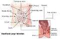
Ascending colon
| Ascending colon | |
|---|---|
 Drawing of colon seen from front (ascending colon coloured blue) | |
 | |
| Details | |
| Precursor | Midgut |
| Artery | Right colic artery |
| Vein | Right colic vein |
| Nerve | Celiac ganglia, vagus[1] |
| Identifiers | |
| Latin | Colon ascendens |
| TA | A05.7.03.002 |
| FMA | 14545 |
The ascending colon is the part of the colon located between the cecum and the transverse colon.
The ascending colon is smaller in caliber than the cecum from where it starts. It passes upward, opposite the colic valve, to the under surface of the right lobe of the liver, on the right of the gall-bladder, where it is lodged in a shallow depression, the colic impression; here it bends abruptly forward and to the left, forming the right colic flexure (hepatic) where it becomes the transverse colon.
It is retained in contact with the posterior wall of the abdomen by the peritoneum, which covers its anterior surface and sides, its posterior surface being connected by loose areolar tissue with the iliacus, quadratus lumborum, aponeurotic origin of transversus abdominis, and with the front of the lower and lateral part of the right kidney.
Sometimes the peritoneum completely invests it, and forms a distinct but narrow mesocolon.
It is in relation, in front, with the convolutions of the ileum and the abdominal walls.
Parasympathetic innervation to the ascending colon is supplied by the vagus nerve. Sympathetic innervation is supplied by the thoracic splanchnic nerves.
Additional images
-

1: Ascending colon
2: Transverse colon
3: Descending colon
4: Sigmoid colon
5: Rectum -
Intestines
-
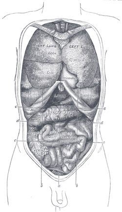
Front view of the thoracic and abdominal viscera.
-

Horizontal disposition of the peritoneum in the lower part of the abdomen.
-
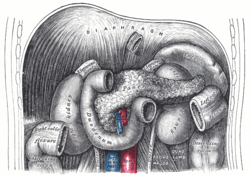
The duodenum and pancreas.
-
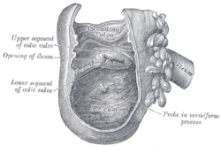
Interior of the cecum and lower end of ascending colon, showing colic valve.
-

Transverse section through the middle of the first lumbar vertebra, showing the relations of the pancreas.
-
The relations of the kidneys from behind.
-

-
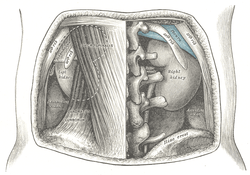
Ascending colon
-
Mesenteric relation of intestines. Deep dissection. Anterior view.
See also
References
This article incorporates text in the public domain from the 20th edition of Gray's Anatomy (1918)
- ↑ Physiology: 6/6ch2/s6ch2_30 - Essentials of Human Physiology
External links
- Anatomy figure: 37:06-08 at Human Anatomy Online, SUNY Downstate Medical Center - "The large intestine."
- largeintestine at The Anatomy Lesson by Wesley Norman (Georgetown University) (cecuminside)