Axillary arch
| Axillary arch | |
|---|---|
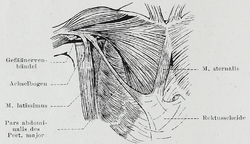 Axillary arch labelled as 'achselbogen'. | |
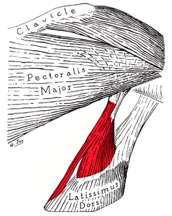 Axillary arch highlighted in red. | |
| Details | |
| Origin | Latissimus dorsi muscle |
| Insertion | Humerus, Pectoralis major |
| Artery | Lateral thoracic artery and subscapular artery |
| Nerve | Lateral pectoral nerve |
| Latin | Arcus axillaris |
The Axillary arch (/ækˈsɪləriː ɑːtʃ/) is a sporadic variant in the musculature associated with the latissimus dorsi muscle in humans. The axillary arch is formed when either a muscular or fascial slip develops extending between the latissimus dorsi and the pectoralis major. The axillary arch shows considerable variation in the exact position of its origin and insertions as well as its pattern of vascularisation and innervation.[1] The axillary arch has several other names including Langer's axillary arch, the muscle of Langer, axillopectoral muscle, Achselbogen and pectorodorsalis muscle.[2][3]
Structure
The axillary arch has a highly variable structure but its defining characteristics are its origin in the latissimus dorsi muscle, its insertion close to or on the antero-superior part of the humerus and that it crosses the neurovascular bundle associated with the axillary nerve from dorsomedial to ventrolateral.[4]
Innervation
As well as its most frequent presentation of innervation from the lateral pectoral nerve instances have been found of innervation from the intercostobrachial nerve, the medial pectoral nerve and the thoracodorsal nerve. [1]
Variations
The extent of the muscularisation of the arch is variable with almost purely fibrous arches also being observed.[5] The exact position of the origin of the axillary arch varies and can be a direct continuation of the latissimus dorsi muscle fibres, originate with the tendinous element of the latissimus dorsi or be a mixture of the two types originating from both the muscle and the tendon.[6]
Insertions are even more variable, Hirtler (2014) cites reported cases occurring at the tendon of the pectoralis major muscle, the pectoralis major muscle, the fascia of the coracobrachialis muscle, the fascia covering the biceps brachii muscle, the long head of the biceps brachii muscle, the coracoid process, the pectoralis minor muscle, the axillary fascia and to the bone at the crest of the greater tubercle of the humerus inserting distal to the insertion of the pectoralis major muscle.[2]
Function
The axillary arch has no apparent function and there is some doubt as to the exact nature of its relationship to structures in other animals.
Prevalence
The general prevalence of the axillary arch is ~7% but this varies considerably amongst populations. In the Turkish population the prevalence is much lower at 1.7% while in the Chinese population it is much higher at 43.8%.[7][8][2]
Clinical relevance
There are several potential clinical consequences of the presence of an axillary arch including confusing the identification and palpation of enlarged or tumorous lymph nodes, the trapping of axillary structures including the axillary nerve and the axillary vein and causing potential problems in axillary surgery or breast reconstruction.[2][9][10] There have also been reported instances of the axillary arch being involved in the development of deep vein thrombosis.[11]
History
The axillary arch muscle was first described by Bugnone in 1783 according to Pitzorno (1912) with Alexander Ramsay describing it as a novel variation during dissections performed in Edinburgh and London around 1793 by his own account of 1813.[12][13] In 1846 Karl Langer wrote up an account of the axillary arch, although he initially only described it as a fibrous arch, and termed it the Achselbogen.[5] Langer later described the presence of a muscular slip associated with the arch in certain cases.[14] Due to these publications Leo Testut used the term arc axillaire de Langer to refer to the muscular form of the arch, an association which has persisted.[15]
Other animals
Possible homologous structures in other species have been identified as the dorsoepitrochlearis muscle, the pectoralis quartus muscle or the panniculus carnosus.[1][2] The dorsoepitrochlearis is an important climbing muscle in monkeys and apes where it has its origin in the tendonous region of the latissimus dorsi and extends down the arm as a superficial muscle.[16]
The panniculus carnosus is a layer of striated muscle deep to the panniculus adiposus.[17] Although absent in hominoids the panniculus carnosus is common in non-hominoid primates and non-primate mammals.[18] In lower mammals the area of the panniculus carnosus can be extensive, almost covering the entire body in the case of the short-beaked echidna.[19] Several human muscles are considered discrete muscles originally part of the panniculus carnosus and some researchers classify the axillary arch as a sporadic vestigial muscle of this type.[20]
See also
References
- 1 2 3 Wilson JT, The Innervation of the Achselbogen Muscle, J Anat Physiol. 1912 Oct; 47(Pt 1): 8–17.
- 1 2 3 4 5 Hirtler L, Langer’s Axillary Arch – Case Presentation and Literature Overview, Austin publishing group, Austin J Anat. 2014;1(4): 1020.
- ↑ Bergman, Ronald. "Axillary Arch". Anatomy Atlases. Retrieved 28 September 2015.
- ↑ Jelev L, Georgiev GP, Surchev L., Axillary arch in human: common morphology and variety. Definition of "clinical" axillary arch and its classification., Ann Anat. 2007;189(5):473-81.
- 1 2 Langer C., Zur Anatomie des Musculus latissimus dorsi., Österr Med Wochenschrift., 1846, 15: 454 - 458.
- ↑ Dharap A., An unusually medial axillary arch muscle., J Anat. 1994, 184: 639-641. PMID 7928652
- ↑ Kalaycioglu A, Gümüsalan Y, Ozan H., Anomalous insertional slip of latissimus dorsi muscle: arcus axillaris., Surg Radiol Anat. 1998; 20: 73-75.
- ↑ Wagenseil F., Muskelbefunde bei Chinesen., Verh. Ges. Phys. Anthrop., Stuttgart, 1927, 2:42–50.
- ↑ Rizk E, Harbaugh K., The muscular axillary arch: an anatomic study and clinical considerations., Neurosurgery. 2008 Oct;63(4 Suppl 2):316-9.
- ↑ Bharambe VK, Arole V, The axillary arch muscle (Langer's muscle): Clinical importance, Med J DY Patil Univ, 2013, 6:327-30.
- ↑ Magee C, Jones C, McIntosh S, Harkin DW., Upper limb deep vein thrombosis due to Langer's axillary arch., J Vasc Surg. 2012 Jan;55(1):234-6.
- ↑ Pitzorno H., Contributo alla morfologia dell'arco ascellare musculare di Langer., Arch Ital Anat Embryol., 1911, 10: 129-144.
- ↑ Ramsay A., An account of unusual conformation of some muscles and vessels., Edinburgh Med. Surg. J., 1812, 8:281-283.
- ↑ Langer C., Zur Anatomie des Musculus latissimus dorsi (Schluss). Österr Med Wochenschrift., 1846, 16: 486-492.
- ↑ Testut L., Les Anomalies Musculaires chez l'Homme Expliquées par l'Anatomie Comparée et leur Importance en Anthropologie., Paris: Masson, 1884.
- ↑ Edwards, William E., The Musculoskeletal Anatomy of the Thorax and Brachium of an Adult Female Chimpanzee,6571st Aeromedical Research Laboratory, New Mexico, 1965.
- ↑ McGrath, J.A.; Eady, R.A.; Pope, F.M. (2004). Rook's Textbook of Dermatology (Seventh Edition). Blackwell Publishing. Page 3.1. ISBN 978-0-632-06429-8.
- ↑ Diogo R, Wood BA, Comparative Anatomy and Phylogeny of Primate Muscles and Human Evolution, CRC Press, 2012. ISBN 9781578087679
- ↑ Griffiths M, Walton DW (Editor), Richardson BJ(Editor), Fauna of Australia Volume 1B, AGPS Canberra, 1989.
- ↑ Bonastre V, Rodríguez-Niedenführ M, Choi D, Sañudo JR, Coexistence of a pectoralis quartus muscle and an unusual axillary arch: case report and review., Clin. Anat. 15:366–370, 2002.