Cherubism
| Cherubism | |
|---|---|
 | |
| A 37-year-old woman exhibiting symptoms of mild cherubism | |
| Classification and external resources | |
| Specialty | gastroenterology |
| ICD-10 | K10.8 |
| ICD-9-CM | 526.89 |
| OMIM | 118400 |
| DiseasesDB | 31217 |
| MedlinePlus | 001234 |
| eMedicine | radio/284 |
| MeSH | D002636 |
Cherubism is a rare genetic disorder that causes prominence in the lower portion in the face. The name is derived from the temporary chubby-cheeked resemblance to putti, often confused with cherubs, in Renaissance paintings.
History
"Cherubism" was first coined and documented in 1933 by Dr. W. A. Jones of Kingston, Ontario, describing a case of three siblings of the same family of Jewish Russian heritage. All that was known at the time was the characteristic swelling pattern and the increase and then regress of bone lesions.[1] By the time the children reached the ages of fifteen, sixteen, and seventeen, the facial deformity had become "grotesque", and in 1943, the children were operated on by the Jones medical team, reducing the hard swelling of their jaws. Four years following the surgeries, there was no reappearance of the swellings.[2]
Presentation
The appearance of people with the disorder is caused by a loss of bone in the mandible which the body replaces with excessive amounts of fibrous tissue. In most cases, the condition fades as the child grows, but in a few even rarer cases the condition continues to deform the affected person's face. Cherubism also causes premature loss of the primary teeth and uneruption of the permanent teeth.
The disease Cherubism is a rare autosomal dominant disease of the maxilla and mandible. Approximately 200 cases have been reported by medical journals with the majority being males. Cherubism is usually first diagnosed around age 7 and continues through puberty and may or may not continue to advance with age.[3] The degrees of Cherubism vary from mild to severe. Osteoclastic and osteoblastic remodeling contributes to the change of normal bone to fibrous tissue and cyst formation. As noted by the name, the patient's face becomes enlarged and disproportionate due to the fibrous tissue and atypical bone formation. The sponge-like bone formations lead to early tooth loss and permanent tooth eruption problems. The disease also affects the orbital area, creating an upturned eye appearance. The cause of cherubism is believed to be traced to a genetic defect resulting from a mutation of the SH3BP2 gene from chromosome 4p16.3.[4] While the disease is rare and painless, the afflicted suffer the emotional trauma of disfigurement. The effects of Cherubism may also interfere with normal jaw motion and speech. Currently, removal of the tissue and bone by surgery is the only treatment available. This disease is also one of the few that unexpectedly stops and regresses.[5] Normal bone remodeling activity may resume after puberty.
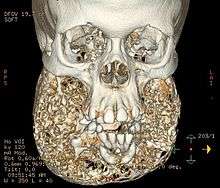
Cherubism is displayed with genetic conformation and when excessive osteoclasts are found in the affected areas of the mandible and maxilla. Large cysts will be present with excessive fibrous areas inside the bone. The fibers and cysts will be found among the trabecula of the Coronoid process, the ramus of mandible, the body of mandible and the maxilla regions. The maxilla will be affected up to and including the orbits and sometimes inside the lower orbits.[6] The maxilla and zygomatic bones are depressed and eyes appear to gaze upward.[7] The maxilla has been found to be more severely affected in most cases than the mandible bone. Some patients found with lower inner orbital growths and cysts may lose vision.
Causes
Cherubism is autosomal dominantly linked, meaning the displayed phenotype is determined by the dominant allele while the normal allele is recessive. One copy of the dominant allele is enough to cause the disorder. Because the disease was found to be dominant the diseased phenotype tends to be seen in every generation at some level of severity. Therefore, afflicted fathers or mothers of children with Cherubism pass the phenotype to both daughters and sons.[5] Cherubism has also been found from the random mutation of a gene in an individual having no family history of the disease. However it is not well understood why males tend to express the disease more frequently. The disease is expressed at a rate of 80 to 100% of all affected, yet children with Cherubism vary in severity in their maxilla and mandible bony lesions.
The cause of Cherubism is believed to be from a mutation of gene of SH3BP2. Cherubism has also been found combined with other genetic disorders including Noonan syndrome, Ramon syndrome, and Fragile X syndrome.[8] Mutations of the SH3BP2 gene are only reported in 75% of Cherubism cases.[4] The mutation of the SH3BP2 gene is believed to increase production of over active proteins from this gene. The SH3BP2 gene is found on the smaller arm of chromosome 4 at position 16.3.[4] The SH3BP2 protein is involved with chemical signaling to immune system cells known as macrophages and B cells.
The effects of SH3BP2 mutations are still under study, but researchers believe that the abnormal protein disrupts critical signaling pathways in cells associated with the maintenance of bone tissue and in some immune system cells. The overactive protein likely causes inflammation in the jaw bones and triggers the production of osteoclasts, which are cells that break down bone tissue during bone remodeling. Osteoclasts also sense the increased inflammation of the mandible and maxilla and are further activated to break down bone structures. Bone loss and inflammation lead to increased fibrous tissue and cyst growth. An excess of these bone-eating cells contributes to the destruction of bone in the upper and lower jaws. A combination of bone loss and inflammation likely underlies the cyst-like growths characteristic of Cherubism.
Diagnosis
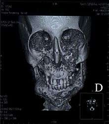
The disease is usually diagnosed when dental abnormalities are found. These abnormalities include premature deciduous teeth and abnormal growth of permanent teeth due to displacement by cysts and lesions. The only definite way to correctly diagnose the disease is by sequence analysis of the SH3BP2 gene. The gene has been found to have missense mutation in exon 9.[5] Initial study of the patient is usually conducted using x-ray and CT scans. Neurofibromatosis may resemble Cherubism and may accompany the disease. Genetic testing is the final diagnosis tool.
Treatment
Because Cherubism changes and improves over time the treatment should be individually determined. Generally moderate cases are watched until they subside or progress into the more severe range. Severe cases may require surgery to eliminate bulk cysts and fibrous growth of the maxilla and mandible. Surgical bone grafting of the cranial facial bones may be successful on some patients. Surgery is preferred for patients ages 5 to 15.[3] Special consideration should be taken when operating on the face to avoid the marginal mandibular branch of the facial nerve as well as the zygomatic branch of the facial nerve. Unintentional damage to these nerves can decrease muscle strength in the face and mandible region. Orthodontic treatment is generally required to avoid permanent dental problems arising from malocclusive bite, misplaced, and unerupted permanent teeth.[3] Orthodontic treatment may be used to erupt permanent teeth that have been unable to descend due to lesions and cysts being in their path of eruption. In patients with orbital issues of diplopia, eye proptosis, and visual loss will require ophthalmologic treatment.[3]
Prognosis
Due to the rarity of the disease, it's difficult to reliably estimate statistics. However, a 2006 study which followed 7 cases over an average of 8.5 years noted that "In general, cherubism does not have a poor prognosis. It has been noted that the condition does not progress beyond puberty. As the patient grows to adulthood, the jawbone lesions tend to resolve, and a progressively more normal jaw configuration is noted.".[9]
Prevention
Because this disease is genetically linked, genetic counseling may be the only way to decrease occurrences of Cherubism. The lack of severe symptoms in the parents may be the cause of failure in recognizing the disorder. The optimal time to be tested for mutations is prior to having children. The disease results from a genetic mutation, and this gene has been found to spontaneously mutate. Therefore, there may be no prevention techniques available.
See also
References
- ↑ Jones, W. A. (1933). "Familial Multilocular Cystic Disease of the Jaws". The American Journal of Cancer. 17 (4): 946. doi:10.1158/ajc.1933.946.
- ↑ Jones, W. A.; Gerrie, J.; Pritchard, J. (1950). "Cherubism--familial fibrous dysplasia of the jaws". The Journal of bone and joint surgery. British volume. 32–B (3): 334–347. PMID 14778852.
- 1 2 3 4 Carroll AL, Sullivan TJ (February 2001). "Orbital involvement in cherubism". Clin. Experiment. Ophthalmol. 29 (1): 38–40. doi:10.1046/j.1442-9071.2001.00363.x. PMID 11272784.
- 1 2 3 Li CY, Yu SF (2006). "A novel mutation in the SH3BP2 gene causes cherubism: case report". BMC Med. Genet. 7: 84. doi:10.1186/1471-2350-7-84. PMC 1764878
 . PMID 17147794.
 - 1 2 3 Tiziani V, Reichenberger E, Buzzo CL, et al. (July 1999). "The gene for cherubism maps to chromosome 4p16". American Journal of Human Genetics. 65 (1): 158–66. doi:10.1086/302456. PMC 1378086. PMID 10364528.
- ↑ Irfan S, Cassels-Brown A, Hayward J, Corrigan A (1997). "Orbital Cherubism". Orbit. 16: 109–112. doi:10.3109/01676839709019125.
- ↑ Emoto Y, Emoto H, Fujle W, Wakakura M (2007). "Uncorrectable Oblique Astigmatism and Impaired Binocular Vision in Case of Orbital Cherubism". Neuro-ophthalmology. 31: 191–5. doi:10.1080/01658100701648553.
- ↑ van Capelle CI, Hogeman PH, van der Sijs-Bos CJ, et al. (September 2007). "Neurofibromatosis presenting with a cherubism phenotype". Eur. J. Pediatr. 166 (9): 905–9. doi:10.1007/s00431-006-0334-6. PMID 17120035.
- ↑ Peñarrocha, Miguel; Bonet, Jaime; Mínguez, Juan Manuel; Bagán, José Vicente; Vera, Francisco; Mínguez, Ignacio (2006-06-01). "Cherubism: a clinical, radiographic, and histopathologic comparison of 7 cases". Journal of Oral and Maxillofacial Surgery: Official Journal of the American Association of Oral and Maxillofacial Surgeons. 64 (6): 924–930. doi:10.1016/j.joms.2006.02.003. ISSN 0278-2391. PMID 16713807.
External links
- "Growing up with cherubism" — personal account on BBC website by Victoria Wright nee Lucas
- Changing Faces A UK-based charity giving support and information to people with disfigurements to the face, hands or body, and their families.
- National Library of Medicine. Cherubism
- GeneReviews/NIH/UW entry on Cherubism
- Imaging of craniofacial fibrous dysplasia Journal of Medical Imaging and Radiation Oncology
- Imaging characteristics of cherubism American Journal of Roentgenology
- Treatment of fibrous dysplasia of the mandible with radical excision and immediate reconstruction: case report The Journal of Craniofacial Surgery