Cover slip
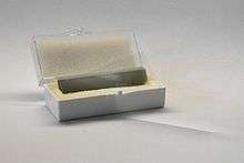
A cover slip, coverslip or cover glass is a thin flat piece of transparent material, usually square or rectangular, about 20 mm (4/5 in) wide and a fraction of a millimetre thick, that is placed over objects for viewing with a microscope. The object is usually held between the cover slip and a somewhat thicker microscope slide, which rests on the microscope's stage or slide holder and provides the physical support for the object and slip.
The main function of the cover slip is to keep solid specimens pressed flat, and liquid samples shaped into a flat layer of even thickness. This is necessary because high-resolution microscopes have a very narrow region within which they focus.
The cover glass often has several other functions. It holds the specimen in place (either by the weight of the cover slip or, in the case of a wet mount, by surface tension) and protects the specimen from dust and accidental contact. It protects the microscope's objective lens from contacting the specimen and vice versa; in oil immersion microscopy or water immersion microscopy the cover slip prevents contact between the immersion liquid and the specimen. The cover slip can be glued to the slide so as to seal off the specimen, retarding dehydration and oxidation of the specimen. Microbial and cell cultures can be grown directly on the cover slip before it is placed on the slide, and specimens may be permanently mounted on the slip instead of on the slide.[1]
Dimensions
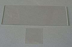
Cover slips are available in a variety of widths, lengths, and thicknesses. They are usually sized so as to fit well inside the boundaries of the microscope slide, which typically measures 25 by 75 mm. Square and round slips are usually 20 mm wide or smaller. Rectangular slips measuring up to 24 by 60 mm are commercially available.[2]
Cover slips are widely available in several standard thicknesses, identified by numbers:[3]
- No. 0 – 0.085 to 0.13 mm thick
- No. 1 – 0.13 to 0.16 mm thick
- No. 1.5 – 0.16 to 0.19 mm thick
- No. 1.5H – 0.17 to 0.18 mm thick**
- No. 2 – 0.19 to 0.23 mm thick
- No. 3 – 0.25 to 0.35 mm thick
- No. 4 – 0.43 to 0.64 mm thick
The thickness of the cover slip is crucially important for high-resolution microscopy. Typical biological microscope objectives are designed for use with No. 1.5 cover slips (0.17 mm thickness).
This is a common statement in the last 30 years with the recommendation to use #1 1/2 cover-slips which are 0.17 mm thick. However the German literature clearly indicates that the so-called "cover-glass thickness" really means the thickness of the cover glass PLUS the thickness of the mount, when the specimen is attached to the slide. In other words, it is the thickness of the cover glass PLUS the thickness of the mount from the underside of the cover glass to the top of the specimen e.g. histological slide. Therefore, the desirable cover slip is a #1 which is approximately 0.13-0.15 mm thick and which allows a little space for the mount. Even the effective "cover glass thickness" of these #1 slips with mount will exceed 0.17 mm.
This is easily confirmed by using a 40x achromatic objective with an NA=0.9 and with a correction collar which is adjusted until the image is crisp-with the aperture diaphragm wide open to keep the NA at 0.9.The effective cover-glass thickness can be read off the scale next to the correction collar. 40x objectives with achromatic and fluorite corrections have NA's less than 0.7 and are more tolerant to deviations from the optimal thickness of 0.17 mm "cover glasses".
The important point is that the recommendation to use #1 1/2 cover slips for routine histological slides is incorrect and #1 slips should be used. Approximately 30 years ago there was a sudden change from #1 to #1 1/2 slips because of the misunderstanding of the designation of the "cover-glass thickness" of 0.17 mm.
If the section, smear or tissue culture is applied directly to the cover slip then a #1 1/2 is required to make the distance from the top of the cover glass to the top of the specimen 0.17 mm. Mount is used to attach the cover glass to the slide but it is not involved in cover glass thickness.[4]
Use of cover slips that deviate from this intended thickness will result in spherical aberration and a reduction in resolution and image intensity. Specialty objectives may be intended for use without coverslips, or may have correction collars that permit a user to accommodate for alternative coverslip thickness.[5]
Sealing
The cover slip is often glued to the microscope slide, in order to seal off the specimen from contamination and decay. A number of sealants are in use, including commercial sealants, laboratory preparations, or even regular clear nail polish, depending on the sample. A solvent-free sealant that can be used for live cell samples is "valap", a mixture of vaseline, lanolin and paraffin in equal parts.[1]
References
- 1 2 Microscopy - Protocols teaching webpage by the Moores Cancer Center, University of California at San Diego. Accessed on 2013-02-07.
- ↑ Coverslip/Coverglass catalog page from a commercial website (Warner Instruments). Accessed on 2010-01-23.
- ↑ GE technical specs from a commercial website (Ted Pella). Accessed on 2010-01-23.
- ↑ Schenk R and Kistler G, Photomicrogphy, London, Chapmam and Hall p38. 1962
- ↑ Michael W. Davidson (2010), Optical Aberrations, chapter in Molecular Expressions website at Florida State University. Last edit 2006-06-15 at 02:39 PM, accessed in 2010-01-12.