Episcleritis
| Episcleritis | |
|---|---|
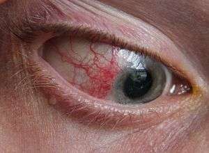 | |
| Eye with Episcleritis | |
| Classification and external resources | |
| Specialty | ophthalmology |
| ICD-10 | H15.1 |
| ICD-9-CM | 379.0 |
| DiseasesDB | 4375 |
| MedlinePlus | 001019 |
| MeSH | D015423 |
Episcleritis is a benign, self-limiting inflammatory disease affecting part of the eye called the episclera. The episclera is a thin layer of tissue that lies between the conjunctiva and the connective tissue layer that forms the white of the eye (sclera). Episcleritis is a common condition, and is characterized by the abrupt onset of mild eye pain and redness.
There are two types of episcleritis, nodular and simple. Nodular episcleritis lesions have raised surface. Simple episcleritis lesions are flat. There are two subtypes. In diffuse simple episcleritis, inflammation is generalized. In sectoral simple episcleritis, the inflammation is restricted to one region.
Most cases of episcleritis have no identifiable cause, although a small fraction of cases are associated with various systemic diseases. Often people with episcleritis experience it recurrently. Treatment focuses on decreasing discomfort, and includes lubricating eye drops. More severe cases may be treated with topical corticosteroids or oral anti-inflammatory medications (NSAIDs).
Signs and symptoms
Symptoms of episcleritis include mild eye pain, redness, and watery eyes.[1] The pain of episcleritis is typically mild, less severe than in scleritis,[2] and may be tender to palpation.[3]
There are two types of episcleritis: the diffuse type, where the redness involves the entire episclera, and the nodular type, where the redness appears more nodular, involving only a small, well-circumscribed area (sectoral).[4] The diffuse type of episcleritis may be less painful than the nodular type. Sometimes, small nodules are present within the episclera, which move slightly over the sclera with gentle pressure.[4]
Discharge is absent with episcleritis, and vision is unaffected.[4] Patients with episcleritis experience far less photophobia than patients with uveitis.[1] Episcleritis does not cause the presence of cells or flare in the anterior chamber of the eye.[1]
Pathophysiology
Most of the time, the cause of episcleritis is never determined (idiopathic). An identifiable cause is discovered in about one third of cases.[5] Several diseases are associated with episcleritis, including systemic vasculitic diseases (polyarteritis nodosa, granulomatosis with polyangiitis), connective tissue diseases (rheumatoid arthritis),[6] rosacea, atopy,[5] gout,[4] and ulcerative colitis.[7] 59 percent of patients with relapsing polychondritis have either episcleritis or scleritis.[8] Rarely, episcleritis may be caused by scleritis.[9]
The redness in the eye associated with episcleritis is due to engorgement of the large episcleral blood vessels, which run in a radial direction from the limbus.[4] Typically, there is no uveitis, or thickening of the sclera.
Diagnosis
The diagnosis of episcleritis is based upon the history and physical examination. The history should be explored for the presence of the diseases associated with episcleritis, and the symptoms they cause, such as rash, arthritis, venereal disease, and recent viral infection.[4] Episcleritis may be differentiated from scleritis by using phenylephrine or neosynephrine eye drops, which causes blanching of the blood vessels in episcleritis, but not in scleritis.[2] A blue color to the sclera suggests scleritis, rather than episcleritis.
After anesthetizing the eye with medication, the conjunctiva may be moved with a cotton swab to observe the location of the enlarged blood vessels.
Treatment
Often, treatment is not necessary, because episcleritis is a self-limiting condition. Artificial tears may be used to help with irritation and discomfort. More severe cases can be treated with either topical corticosteroids or oral non-steroidal anti-inflammatory drugs.[5]
Ketorolac, a topical NSAID, may be used, but it is not more effective than artificial tears and it causes more side effects.[10]
Epidemiology
Episcleritis is a common disease,[11] and its exact prevalence and incidence are unknown. It typically affects young adults,[11] and may be more common in women.
Prognosis
Episcleritis is a benign, self-limiting condition, meaning patients recover without any treatment. Most cases of episcleritis resolve within 7–10 days.[1] The nodular type is more aggressive and takes longer to resolve.[1] Although rare, some cases may progress to scleritis.[12] However, in general, episcleritis does not cause complications in the eye.[12] Smoking tobacco delays the response to treatment in patients with episcleritis.[13]
References
- 1 2 3 4 5 Heath, Greg (10 February 2010). "The episclera, sclera and conjunctiva An overview of relevant ocular anatomy" (PDF). OT: 36–42. Retrieved 30 November 2012.
- 1 2 Goldman, Lee (2011). Goldman's Cecil Medicine (24th ed.). Philadelphia: Elsevier Saunders. p. 2440. ISBN 1437727883.
- ↑ Chumley H.; Usatine RP; Smith MA; Chumley H; Mayeaux, Jr. E; Tysinger J (2009). The Color Atlas of Family Medicine: Chapter 16. Scleritis and Episcleritis. (2nd ed.). New York: McGraw-Hill Education Medical. ISBN 978-0071769648.
- 1 2 3 4 5 6 Kunimoto, Derek; Kunal Kanitkar; Mary Makar (2004). The Wills eye manual: office and emergency room diagnosis and treatment of eye disease. (4 ed.). Philadelphia, PA: Lippincott Williams & Wilkins. pp. 99–100. ISBN 978-0781742078.
- 1 2 3 Yanoff, Myron; Jay S. Duker (2008). Ophthalmology (3rd ed.). Edinburgh: Mosby. pp. 255–261. ISBN 978-0323057516.
- ↑ Watson, PG; Hayreh, SS (March 1976). "Scleritis and episcleritis.". The British journal of ophthalmology. 60 (3): 163–91. doi:10.1136/bjo.60.3.163. PMID 1268179.
- ↑ Langholz, E. (March 2010). "Review: Current trends in inflammatory bowel disease: the natural history". Therapeutic Advances in Gastroenterology. 3 (2): 77–86. doi:10.1177/1756283X10361304. PMC 3002570
 . PMID 21180592.
 - ↑ Sabatine, Marc S. (2011). Pocket Medicine (4th ed.). Philadelphia: Wolters Kluwer Health/Lippincott Williams & Wilkins. p. 8-4. ISBN 1608319059.
- ↑ "Episcleritis: MedlinePlus Medical Encyclopedia". Bethesda, MD: United States National Library of Medicine. Retrieved 20 June 2010.
- ↑ Williams, CP; Browning, AC; Sleep, TJ; Webber, SK; McGill, JI (July 2005). "A randomised, double-blind trial of topical ketorolac vs artificial tears for the treatment of episcleritis.". Eye (London, England). 19 (7): 739–42. doi:10.1038/sj.eye.6701632. PMID 15359265.
- 1 2 Levsky M.E.; DeFlorio P. (2010). Atlas of emergency medicine: Chapter 2 Ophthalmologic Conditions. (3rd ed.). New York: McGraw-Hill Professional. ISBN 978-0071496186.
- 1 2 Jabs, Douglas A.; Mudun, Abdulbaki; Dunn, J.P.; Marsh, Marta J. (Oct 2000). "Episcleritis and scleritis: clinical features and treatment results". American Journal of Ophthalmology. 130 (4): 469–476. doi:10.1016/S0002-9394(00)00710-8. PMID 11024419. Retrieved 30 November 2012.
- ↑ Boonman, Z F H M; de Keizer, R J W; Watson, P G (September 2005). "Smoking delays the response to treatment in episcleritis and scleritis". Eye. 19 (9): 949–955. doi:10.1038/sj.eye.6701731. PMID 15543188.
- Leibowitz, HM; Hyndiuk, RA; Lindsey, C; Rosenthal, AL (December 1984). "Fluorometholone acetate: clinical evaluation in the treatment of external ocular inflammation.". Annals of Ophthalmology. 16 (12): 1110–5. PMID 6398026.
- McGavin, DD; Williamson, J; Forrester, JV; Foulds, WS; Buchanan, WW; Dick, WC; Lee, P; MacSween, RN; Whaley, K (March 1976). "Episcleritis and scleritis. A study of their clinical manifestations and association with rheumatoid arthritis.". The British journal of ophthalmology. 60 (3): 192–226. doi:10.1136/bjo.60.3.192. PMID 1268180.
- Sainz de la Maza, M; Jabbur, NS; Foster, CS (February 1994). "Severity of scleritis and episcleritis.". Ophthalmology. 101 (2): 389–96. doi:10.1016/s0161-6420(94)31325-x. PMID 8115160.
- Watson P. Diseases of the sclera and episclera. In: Tasman W, Jaeger EA, eds. Duane’s Ophthalmology. 15th ed. Philadelphia, Pa: Lippincott Williams & Wilkins; 2009:chap 23.