Fordyce spots
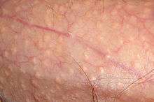
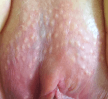
Fordyce spots (also termed Fordyce granules[1] or Fordyce disease)[1][2] are visible sebaceous glands that are present in most individuals. They appear on the genitals and/or on the face and in the mouth. They appear as small, painless, raised, pale, red or white spots or bumps 1 to 3 mm in diameter that may appear on the scrotum, shaft of the penis or on the labia, as well as the inner surface (retromolar mucosa) and vermilion border of the lips of the face. They are not associated with any disease or illness, nor are they infectious but rather they represent a natural occurrence on the body. No treatment is therefore required, unless the individual has cosmetic concerns. Persons with this condition sometimes consult a dermatologist because they are worried they may have a sexually transmitted disease (especially genital warts) or some form of cancer.[3]
Classification
Sebaceous glands are normal adnexal structures of the dermis but may also be found ectopically within the mouth, where they are referred to as Oral Fordyce Granules or ectopic sebaceous glands. On the foreskin they are called Tyson's glands,[4] not to be confused with hirsuties coronae glandis.[5]
When they appear on the penis, they are also called penile sebaceous glands.[6]
When seen as a streak of individual glands along the interface between the skin of the lip and the vermilion border, the terms Fox–Fordyce disease and Fordyce's condition have been used.
Signs and symptoms
On the shaft of the penis, Fordyce spots are more visible when the skin is stretched, and may only be noticeable during an erection.[6] The spots can also appear on the skin of the scrotum.[6]
Oral Fordyce granules appear as rice-like granules, white or yellow-white in color. They are painless papules (small bumps), about 1–3 mm in greatest dimension. The most common site is along the line between the vermilion border and the oral mucosa of the upper lip, or on the buccal mucosa (inside the cheeks) in the commissural region,[1] often bilaterally. They may also occur on the mandibular retromolar pad and tonsillar areas, but any oral surface may be involved. There is no surrounding mucosal change. Some patients will have hundreds of granules while most have only one or two.
Occasionally, several adjacent glands will coalesce into a larger cauliflower-like cluster similar to sebaceous hyperplasia of the skin. In such an instance, it may be difficult to determine whether or not to diagnose the lesion as sebaceous hyperplasia or sebaceous adenoma. The distinction may be moot because both entities have the same treatment, although the adenoma has a greater growth potential. Sebaceous carcinoma of the oral cavity has been reported, presumably arising from Fordyce granules or hyperplastic foci of sebaceous glands.
In some persons with Fordyce spots, the glands express a thick, chalky discharge when squeezed.[6]
Causes
Normally, sebaceous glands are only found in association with a hair follicle.
They appear to be more obvious in people with greasy skin types, with some rheumatic disorders, and in Hereditary nonpolyposis colorectal cancer.[1] In the latter, the most common site for Fordyce spots is the lower gingiva (gums) and vestibular mucosa.[1]
Diagnosis
Large numbers of lobules coalescing into a definitely elevated mass may be called benign sebaceous hyperplasia, and occasional small keratin-filled pseudocysts may be seen and must be differentiated from epidermoid cyst or dermoid cyst with sebaceous adnexa. The pathologist must be careful to differentiate such lesions from salivary neoplasms with sebaceous cells, such as sebaceous lymphadenoma and sebaceous adenoma, and their malignant counterparts sebaceous lymphadenocarcinoma and sebaceous carcinoma.
Oral Fordyce granules are usually not biopsied because they are readily diagnosed clinically, but they are often seen as incidental findings of mucosal biopsies of the buccal, labial and retromolar mucosa. The granules are similar to normal sebaceous glands of the skin but lack hair follicles and almost always lack a ductal communication with the surface. The glands are located just beneath the overlying epithelium and often produce a local elevation of the epithelium. Individual sebaceous cells are large, with central dark nuclei and abundant foamy cytoplasm. The surrounding stroma may contain occasional chronic inflammatory cells because of trauma with adjacent teeth.
Prognosis
Fordyce spots are completely benign[1] and require no treatment. Often their presence is considered normal anatomic variance rather than a true medical condition.
Treatment
Vaporising laser treatments such as CO2 laser[7] or electro desiccation have been used with some success in diminishing the appearance of this condition if they are of cosmetic concern, despite the fact that most doctors consider this a normal physiological phenomenon and advise against treatment.[8]
Success varies per patient, but some have found relief from pulsed dye lasers,[9] a laser normally used to treat sebaceous gland hyperplasia,[10] which is similar to Fordyce spots. Treatment with pulsed dye lasers is expensive, but may be less likely to scar than other methods.[10]
A surgical treatment using a Micro-punch[11] technique has shown to be an effective method that achieved very satisfactory functional and cosmetic results in patient that have multiple and/or very prominent fordyce spots.
No treatment is required for oral Fordyce granules, except for cosmetic removal of labial lesions if the individual wishes it. Inflamed glands can be treated topically with clindamycin. When surgically excised they will not recur. Neoplastic transformation is very rare but has been reported.
Epidemiology
This variation of normal anatomy is seen in the majority of adults. It is estimated about 80% of people have oral Fordyce spots,[1] but seldom are granules found in large numbers. They are not usually visible in children, and tend to appear at about age 3, then increasing during puberty and become more obvious in later adulthood.[1] They are more prominent in males.[1]
History
They are named after an American dermatologist, John Addison Fordyce.[12]
References
- 1 2 3 4 5 6 7 8 9 Scully C (2013). Oral and maxillofacial medicine : the basis of diagnosis and treatment (3rd ed.). Edinburgh: Churchill Livingstone. pp. 170, 392. ISBN 978-0-7020-4948-4.
- ↑ James, William; Berger, Timothy; Elston, Dirk (2005). Andrews' Diseases of the Skin: Clinical Dermatology. (10th ed.). Saunders. ISBN 0-7216-2921-0.
- ↑ Palo Alto Medical Foundation Bettina McAdoo , M.D. Retrieved June 24, 2006.
- ↑ derm/395 at eMedicine
- ↑ Khoo LS, Cheong WK (July 1995). "Common genital dermatoses in male patients attending a public sexually transmitted disease clinic aka(DC) in Singapore". Annals of the Academy of Medicine, Singapore. 24 (4): 505–9. PMID 8849177.
- 1 2 3 4 Rane V, Read T (May 2013). "Penile appearance, lumps and bumps". Australian Family Physician. 42 (5): 270–4. PMID 23781523.
- ↑ Ocampo-Candiani J, Villarreal-Rodríguez A, Quiñones-Fernández AG, Herz-Ruelas ME, Ruíz-Esparza J (August 2003). "Treatment of Fordyce spots with CO2 laser". Dermatologic Surgery. 29 (8): 869–71. doi:10.1046/j.1524-4725.2003.29236.x. PMID 12859392.
- ↑ Nordqvist, Christian (February 27, 2013). "What Are Fordyce Spots? What Causes Fordyce Spots?". Medical News Today.
- ↑ http://sebaceous.proboards42.com/index.cgi?board=real&action=display&thread=249&page=5[]
- 1 2 Schönermark MP, Schmidt C, Raulin C (1997). "Treatment of sebaceous gland hyperplasia with the pulsed dye laser". Lasers in Surgery and Medicine. 21 (4): 313–6. doi:10.1002/(SICI)1096-9101(1997)21:4<313::AID-LSM1>3.0.CO;2-T. PMID 9328977.
- ↑ Pallua, N.; Stromps, J. P. (January 2013). "Micro-punch technique for treatment of Fordyce spots: A surgical approach for an unpleasant condition of the male genital". Journal of Plastic, Reconstructive & Aesthetic Surgery. 66 (1): e8–e11. doi:10.1016/j.bjps.2012.08.039. PMID 23026471.
- ↑ Fordyce first described this condition in 1896.synd/1510 at Who Named It?
Further reading
- Fordyce J. A peculiar affection of the mucous membrane of the lip and oral cavity. J Cutan Genito-Urin Dis 1896; 14:413-419.
- Miles AEW. Sebaceous glands in the lip and cheek mucosa of man. Br Dent J 1958; 105:235-239.
- Miller AS, McCrea MW (September 1968). "Sebaceous gland adenoma of the buccal mucosa: report of case". Journal of Oral Surgery. 26 (9): 593–5. PMID 4299381.
- Gorsky M, Buchner A, Fundoianu-Dayan D, Cohen C (August 1986). "Fordyce's granules in the oral mucosa of adult Israeli Jews". Community Dentistry and Oral Epidemiology. 14 (4): 231–2. doi:10.1111/j.1600-0528.1986.tb01541.x. PMID 3461911.
- Rhodus NL (1986). "An actively secreting Fordyce granule. A case report". Clinical Preventive Dentistry. 8 (2): 24–6. PMID 3486083.
- Ellis GL, Auclair PL, Gnepp DR. Surgical pathology of the salivary glands. Philadelphia; W.B. Saunders, 1991.
- Miller ML, Harford RR, Yeager JK (October 1995). "Fox-Fordyce disease treated with topical clindamycin solution". Archives of Dermatology. 131 (10): 1112–3. doi:10.1001/archderm.1995.01690220018003. PMID 7574824.
- Abuzeid M, Gangopadhyay K, Rayappa CS, Antonios JI (May 1996). "Intraoral sebaceous carcinoma". The Journal of Laryngology and Otology. 110 (5): 500–2. doi:10.1017/s0022215100134103. PMID 8762330.