Hepatitis delta virus ribozyme
| Hepatitis delta virus ribozyme | |
|---|---|
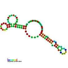 | |
| Predicted secondary structure and sequence conservation of HDV ribozyme | |
| Identifiers | |
| Symbol | HDV_ribozyme |
| Rfam | RF00094 |
| Other data | |
| RNA type | Gene; ribozyme |
| Domain(s) | Viruses |
| SO | 0000374 |

The hepatitis delta virus (HDV) ribozyme is a non-coding RNA found in the hepatitis delta virus that is necessary for viral replication and is thought to be the only catalytic RNA known to be required for viability of a human pathogen. The ribozyme acts to process the RNA transcripts to unit lengths in a self-cleavage reaction. The ribozyme is found to be active in vivo in the absence of any protein factors and is the fastest known naturally occurring self-cleaving RNA.[2]
The crystal structure of this ribozyme has been solved using X-ray crystallography and shows five helical segments connected by a double pseudoknot.[1]
In addition to the sense (genomic version), all HDV viruses also have an anti-genomic version of the HDV ribozyme.[3] This version is not the exact complementary sequence but adopts the same structure as the sense (genomic) strand. The only "significant" differences between the two are a small bulge in P4 stem and a shorter J4/2 junction.
The hepatitis delta virus ribozyme is structurally and biochemically related to the Mammalian CPEB3 ribozyme. Unrelated sequences with high similarity to the HDV ribozyme have evolved through convergent evolution in some retrotransposons (e.g. in the R2 RNA element in insects and in the L1Tc and probably other retrotransposons in trypanosomatids).[4][5][6][7][8]
General Acid Base Chemistry
Similar to the hairpin ribozyme, the HDV ribozyme functions by acid-base catalysis. The functional groups within the RNA have pH values that allow them to interchange between the acidic and basic states. This behavior shows that the HDV ribozyme must have some form of functional groups that has neutral like pH around 6 to 7. Within RNA, typical pKa values for the free nucleosides are around 3.5 to 4.2, a lower pKa acidic value that would unlikely become basic. However, due to the shifted pKa values that causes the acid-base catalysis, there is a likelihood that it may be caused form the structural environment within the nucleoside.[9][10][11][12]
Within the HDV Ribozyme, there is a network of hydrogen bonds to the cytosine that is able to stabilize the protonated form of the cytosine. It will allow the donation and acceptance of the proton at some during the catalysis.[13] In a set of experiments, a point mutations was done to the critical cytosine. Afterwards there was a drop activity and was partially restored when imidazole was added.
With support data, the current model of cytosine acting as a general acid within the reaction to donate a proton to the 5’-bridging oxygen (See figure)
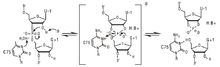
. There is also a metal ion that coordinates near the ribozyme active site to abstract a proton from the 2’-hydroxyl nucleophile (37).[14]
HDV ribozyme has been the subject of many investigations over the years. It is now accepted that HDV ribozyme uses two distinct strategies to catalyze the cleavage of a specific phosphodiester bond.[15] It was recently shown that a Mg2+ ion interacts with the 2’-OH of the scissile nucleotide.[16] This metal is thought to act as a lewis acid which activate the 2’-OH for a nucleophilic attack on the scissile phosphate. C75 is thought to act as a general acid which donates a proton to the 5’ leaving oxygen resulting in the cleavage of the phosphodiester bond. For efficient catalysis, the pKa of C75 needs to be shifted towards neutrality which was confirmed using Raman crystallogphy.[17] It is known that HDV is still active in the absence of divalent metals which leads to the thinking that there is a second catalytic strategy that the ribozyme uses for cleavge. In that case, a water molecule might be deprotonating the 2’-OH rather that Mg2+.
Participation of upstream RNA in regulating catalytic activity of HDV ribozyme
As limited by the rapid self-cleaving nature of HDV ribozyme, the previous ribonuclease experiments were performed on the 3' product of self-cleavage rather than the precursor.[18] However, flanking sequence is known to participate in regulating the self-cleavage activity of HDV ribozyme.[19][20][21] Therefore, the upstream sequence 5' to the self-cleavage site has been incorporated to study the resultant self-cleavage activity of the HDV ribozyme.[19] Two alternative structures have been identified.
The first inhibitory structure is folded by an extended transcript (i.e. -30/99 transcript, coordinates are referenced against the self-cleavage site) spanning from 30 nt upstream of the cleavage site to 15 nt downstream of the 3'-end.[19] The flanking sequence sequesters the ribozyme in a kinetic trap during transcription and results in the extremely diminished self-cleavage rate.[19] This self-cleavage-preventing structure includes 3 alternative stems: Alt1, Alt2 and Alt3, which disrupt the active conformation. Alt1 is a 10-bp Long-Range-Interaction formed by an inhibitory upstream stretch (-25/-15 nt) and the downstream stretch (76/86 nt).[19] The Alt1 disrupts the stem P2 in the active conformation wherein P2 is proposed to have an activating role for both genomic and antigenomic ribozyme.[19][22][23] Alt2 is an interaction between upstream flanking sequence and the ribozyme, and Alt3 is a nonnative ribozyme-ribozyme interaction.[19] The secondary structure of this inhibitory conformation is supported by various experimental approaches.[19] First, direct probing via ribonucleases was performed and the subsequent modeling via mfold 3.0 using constraints from the probing results agrees with the proposed structure.[19] Second, a series of DNA oligomer complementary to different regions of AS1/2 were used to rescue the ribozyme activity; the results confirms the inhibitory roles of AS1/2.[19] Third, mutational analysis introduces single/double mutations outside the ribozyme to ensure the observed ribozyme activity is directly associated with the stability of the Alt1.[19] The stability of AS1 is found to be inversely related to the self-cleavage activity[26].
The second permissive structure enables the HDV ribozyme to self-cleave co-transcriptionally and this structure further includes the -54/-18 nt portion of the RNA transcript.[19] The upstream inhibitory -24/-15 stretch from the aforementioned inhibitory conformation is now sequestered in a hairpin P(-1) located upstream of the cleavage site.[19][24][25] The P(-1) motif, however, is only found in the genomic sequence, which may be correlated with the phenomenon that genomic HDV RNA copies are more abundant in the infected liver cells.[19][26] Experimental evidence also supports this alternative structure. First, structural mapping via ribonuclease is used to probe the -54/-1 fragment instead of the whole precursor transcript due to the fast-cleaving nature of this structure, which agrees with the local hairpin P(-1) (between -54/-40 and -18/-30 nt).[19] Secondly, evolutionary conservation is found in P(-1) and the linking region between P(-1) and P1 among 21 genomic HDV RNA isolates.[19]
References
- 1 2 Ferré-D'Amaré AR, Zhou K, Doudna JA (1998). "Crystal structure of a hepatitis delta virus ribozyme". Nature. 395 (6702): 567–74. doi:10.1038/26912. PMID 9783582.
- ↑ Kuo, MY; Sharmeen L; Dinter-Gottlieb G; Taylor J (1988). "Characterization of self-cleaving RNA sequences on the genome and antigenome of human hepatitis delta virus". J Virol. 62 (12): 4439–4444. PMC 253552
 . PMID 3184270.
 - ↑ Chen PJ, Kalpana G, Goldberg J, et al. (November 1986). "Structure and replication of the genome of the hepatitis delta virus". Proc. Natl. Acad. Sci. U.S.A. 83 (22): 8774–8. doi:10.1073/pnas.83.22.8774. PMC 387014. PMID 2430299.
- ↑ Eickbush, DG; Eickbush, TH (July 2010). "R2 Retrotransposons Encode a Self-Cleaving Ribozyme for Processing from an rRNA Cotranscript". Molecular and Cellular Biology. 30 (13): 3142–50. doi:10.1128/MCB.00300-10. PMC 2897577. PMID 20421411.
- ↑ Webb CH, Riccitelli NJ, Ruminski DJ, Lupták A (November 2009). "Widespread occurrence of self-cleaving ribozymes". Science. 326 (5955): 953. doi:10.1126/science.1178084. PMC 3159031. PMID 19965505.
- ↑ Webb CH, Lupták A (2011). "HDV-like self-cleaving ribozymes". RNA Biol. 8 (5): 719–27. doi:10.4161/rna.8.5.16226. PMC 3256349. PMID 21734469.
- ↑ Sánchez-Luque, FJ; López MC; Macias F; Alonso C; Thomas MC (2011-06-30). "Identification of an hepatitis delta virus-like ribozyme at the mRNA 5′-end of the L1Tc retrotransposon from Trypanosoma cruzi". Nucleic Acids Res. 39 (18): 8065–8077. doi:10.1093/nar/gkr478. PMC 3185411. PMID 21724615.
- ↑ Sánchez-Luque, FJ; López MC; Macias F; Alonso C; Thomas MC (2012-01-01). "Pr77 and L1TcRz. A dual system within the 5′-end of L1Tc retrotransposon, internal promoter and HDV-like ribozyme". Mob Genet Elements. 2 (1): 1–7. doi:10.4161/mge.19233. PMC 3383444. PMID 22754746.
- ↑ Rajagopal, Ponni; Feigon, Juli (22 June 1989). "Triple-strand formation in the homopurine:homopyrimidine DNA oligonucleotides d(G-A)4 and d(T-C)4". Nature. 339 (6226): 637–640. doi:10.1038/339637a0.
- ↑ Sklená\r̆, Vladmír; Felgon, Juli (28 June 1990). "Formation of a stable triplex from a single DNA strand". Nature. 345 (6278): 836–838. doi:10.1038/345836a0.
- ↑ Connell, G.; Yarus, M (20 May 1994). "RNAs with dual specificity and dual RNAs with similar specificity". Science. 264 (5162): 1137–1141. doi:10.1126/science.7513905.
- ↑ Legault, Pascale; Pardi, Arthur (September 1994). "In situ Probing of Adenine Protonation in RNA by 13C NMR". Journal of the American Chemical Society. 116 (18): 8390–8391. doi:10.1021/ja00097a066.
- ↑ Doudna, Jennifer A.; Ferré-D'Amaré, Adrian R.; Zhou, Kaihong (8 October 1998). "Crystal structure of a hepatitis delta virus ribozyme". Nature. 395 (6702): 567–574. doi:10.1038/26912. PMID 9783582.
- ↑ Nakano, Shu-ichi; Proctor, David J.; Bevilacqua, Philip C. (October 2001). "Mechanistic Characterization of the HDV Genomic Ribozyme: Assessing the Catalytic and Structural Contributions of Divalent Metal Ions within a Multichannel Reaction Mechanism". Biochemistry. 40 (40): 12022–12038. doi:10.1021/bi011253n.
- ↑ Golden, B. L. (2011). "Two Distinct Catalytic Strategies in the Hepatitis Delta Virus Ribozyme Cleavage Reaction". Biochemistry. 50: 9424–9433. doi:10.1021/bi201157t.
- ↑ Chen, J.-H.; et al. (2010). "A 1.9 Å Crystal Structure of the HDV Ribozyme Precleavage Suggests both Lewis Acid and General Acid Mechanisms Contribute to Phosphodiester Cleavage". Biochemistry. 49: 6508–6518. doi:10.1021/bi100670p.
- ↑ Gong, B.; et al. (2007). "Direct Measurement of a pKa near Neutrality for the Catalytic Cytosine in the Genomic HDV Ribozyme Using Raman Crystallography". Journal of the American Chemical Society. 129: 13335–13342. doi:10.1021/ja0743893.
- ↑ Rosenstein, SP (1991). "Evidence that genomic and antigenomic RNA self-cleaving elements from hepatitis delta virus have similar secondary structures". Nucleic Acids Res. 19 (19): 5409–16. doi:10.1093/nar/19.19.5409.
- 1 2 3 4 5 6 7 8 9 10 11 12 13 14 15 16 Chadalavada, D.; Knudsen, S.; Nakano, S.; Bevilacqua, P. (2000). "A role for upstream RNA structure in facilitating the catalytic fold of the genomic hepatitis delta virus ribozyme". Journal of Molecular Biology. 301 (2): 349–367. doi:10.1006/jmbi.2000.3953.
- ↑ Perrotta, A. T.; Been, M. D. (1990). "The self-cleaving domain from the genomic RNA of hepatitis delta virus: sequence requirements and the effects of denaturant". Nucleic Acids Res. 18: 6821–6827. doi:10.1093/nar/18.23.6821.
- ↑ Perrotta, A.; Been, M. (1991). "A pseudoknot-like structure required for efficient self-cleavage of hepatitis delta-virus RNA". Nature. 350 (6317): 434–436. doi:10.1038/350434a0.
- ↑ Matysiak, M.; Wrzesinski, J.; Ciesiolka, J. (1999). "Sequential folding of the genomic ribozyme of the hepatitis delta virus: structural analysis of RNA transcription intermediates". J. Mol. Biol. 291: 283–294. doi:10.1006/jmbi.1999.2955.
- ↑ Perrotta, A.T.; Nikiforova, O.; Been, M.D. (1999). "A conserved bulged adenosine in a peripheral duplex of the antigenomic HDV self-cleaving RNA reduces kinetic trapping of inactive conformations". Nucl. Acids Res. 27: 795–802. doi:10.1093/nar/27.3.795.
- ↑ Mathews, D.; Sabina, J.; Zuker, M.; Turner, D. (1999). "Expanded sequence dependence of thermodynamic parameters improves prediction of RNA secondary structure". J. Mol. Biol. 288: 911–940. doi:10.1006/jmbi.1999.2700. PMID 10329189.
- ↑ Zuker, M., Mathews, D., and Turner, D. (1999) Algorithms and thermodynamics for RNA secondary structure prediction practical guide in RNA Biochemistry and Biotechnology, J.B.B.R.C. Clark (Ed.), NATO ASI Series, Kluwer Academic Publishers, Dordrecht, the Netherlands.
- ↑ Chen, P.J., Kalpana, G., Goldberg, J., Mason, W., Werner, B., Gerin, J., Taylor, J. Structure and replication of the genome of the hepatitis delta virus. Proc. Natl Acad. Sci. USA 1986; 83 : 8774–8778