Inflammatory fibroid polyp
| Inflammatory fibroid polyp | |
|---|---|
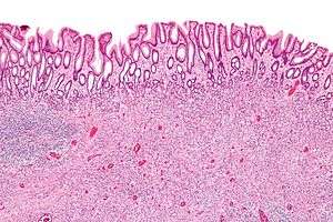 | |
| Micrograph of an inflammatory fibroid polyp. H&E stain. | |
| Classification and external resources |
Inflammatory fibroid polyp, abbreviated IFP, is a benign abnormal growth of tissue projecting into the lumen of the gastrointestinal tract.
Pathology
IFPs consist of spindle cells that are concentrically arranged around blood vessels and have inflammation, especially eosinophils.[1] They may have leiomyoma/schwannoma-like areas with nuclear palisading.
They typically stain with CD34[1] and vimentin,[2] and, generally, do not stain with CD117 and S100.
The endoscopic differential diagnosis includes other benign, pre-malignant and malignant gastrointestinal polyps.
Morphological differential diagnosis
Additional images
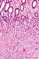 High mag.
See also
Internal links
References
- 1 2 Daum, O.; Hatlova, J.; Mandys, V.; Grossmann, P.; Mukensnabl, P.; Benes, Z.; Michal, M. (May 2010). "Comparison of morphological, immunohistochemical, and molecular genetic features of inflammatory fibroid polyps (Vanek's tumors).". Virchows Arch. 456 (5): 491–7. doi:10.1007/s00428-010-0914-8. PMID 20393746.
- ↑ Kolodziejczyk, P.; Yao, T.; Tsuneyoshi, M. (Nov 1993). "Inflammatory fibroid polyp of the stomach. A special reference to an immunohistochemical profile of 42 cases.". Am J Surg Pathol. 17 (11): 1159–68. doi:10.1097/00000478-199311000-00009. PMID 8214261.
This article is issued from Wikipedia - version of the 1/16/2015. The text is available under the Creative Commons Attribution/Share Alike but additional terms may apply for the media files.