LE cell
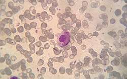
Microphotograph of a macrophage that has phagocytized a lymphocyte (LE cell). May Grünwald Giemsa stain.
An LE cell is a neutrophil or macrophage that has phagocytized (engulfed) the denatured nuclear material of another cell.[1] The denatured material is an absorbed hematoxylin body (also called an LE body).[2]
They are a characteristic of lupus erythematosus,[3] but also found in similar connective tissue disorders.
The LE cell was discovered in bone marrow in 1948 by Malcolm McCallum Hargraves (1903–1982), a Physician and Practicing Histologist at the Mayo Clinic et al.[4]
Classically, the LE cell is analyzed microscopically, but it is also possible to investigate this phenomenon by flow cytometry.[5]
References
- ↑ http://www.merriam-webster.com/medical/le%20cell
- ↑ similima.com > Autoimmunity By Muhammed Muneer. Retrieved Mars 2011
- ↑ http://www.drugsfreelist.com/health_articles/4002/LE_Cell_Test__Lupus_erythematosus_test__
- ↑ Hargraves M, Richmond H, Morton R. Presentation of two bone marrow components, the tart cell and the LE cell. Mayo Clin Proc 1948;27:25–28.
- ↑ Böhm, Ingrid (1 January 2004). "Flow Cytometric Analysis of the LE Cell Phenomenon". Autoimmunity. 37 (1): 37–44. doi:10.1080/08916930310001630325. PMID 15115310.
This article is issued from Wikipedia - version of the 5/9/2016. The text is available under the Creative Commons Attribution/Share Alike but additional terms may apply for the media files.