Lupus nephritis
| Lupus nephritis | |
|---|---|
|
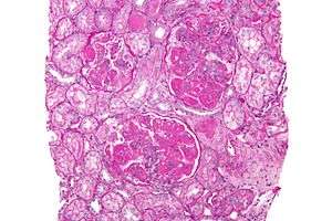
Micrograph of diffuse proliferative lupus nephritis showing increased mesangial matrix and mesangial hypercellularity. Kidney biopsy. PAS stain. | |
| Classification and external resources | |
| Specialty | urology |
| ICD-10 | M32.1+N08.5* |
| ICD-9-CM | 583.81 |
| MedlinePlus | 000481 |
| eMedicine | med/1597 |
| MeSH | D008181 |
Lupus nephritis (also known as SLE nephritis)[1] is an inflammation of the kidneys caused by systemic lupus erythematosus (SLE), an autoimmune disease.[2] It is a type of glomerulonephritis in which the glomeruli become inflamed. As the result of SLE, the cause of glomerulonephritis is said to be secondary and has a different pattern and outcome from conditions with a primary cause originating in the kidney.[3][4]
Signs and symptoms
General symptoms of lupus nephritis include[4][5]
Class I disease (minimal mesangial glomerulonephritis) in its histology has a normal appearance under a light microscope, but mesangial deposits are visible under an electron microscope. At this stage urinalysis is normal.[6]
Class II disease (mesangial proliferative glomerulonephritis) is noted by mesangial hypercellularity and matrix expansion. Microscopic haematuria with or without proteinuria may be seen. Hypertension, nephrotic syndrome, and acute kidney insufficiency are very rare at this stage.[6]
Class III disease (focal glomerulonephritis) is indicated by sclerotic lesions involving less than 50% of the glomeruli, which can be segmental or global, and active or chronic, with endocapillary or extracapillary proliferative lesions. Under the electron microscopy, subendothelial deposits are noted, and some mesangial changes may be present. Immunofluorescence reveals positively for IgG, IgA, IgM, C3, and C1q. Clinically, haematuria and proteinuria are present, with or without nephrotic syndrome, hypertension, and elevated serum creatinine.[6]
Class IV disease (diffuse proliferative nephritis) is both the most severe, and the most common subtype. More than 50% of glomeruli are involved. Lesions can be segmental or global, and active or chronic, with endocapillary or extracapillary proliferative lesions. Under electron microscopy, subendothelial deposits are noted, and some mesangial changes may be present. Clinically, haematuria and proteinuria are present, frequently with nephrotic syndrome, hypertension, hypocomplementemia, elevated anti-dsDNA titres and elevated serum creatinine.[6]
Class V disease (membranous glomerulonephritis) is characterized by diffuse thickening of the glomerular capillary wall (segmentally or globally), with diffuse membrane thickening, and subepithelial deposits seen under the electron microscope. Clinically, stage V presents with signs of nephrotic syndrome. Microscopic haematuria and hypertension may also been seen. Stage V also can also lead to thrombotic complications such as renal vein thromboses or pulmonary emboli.[6]
A final Class is included by most practitioners, Class VI, or advanced sclerosing lupus nephritis.[7] It is represented by global sclerosis involving more than 90% of glomeruli, and represents healing of prior inflammatory injury. Active glomerulonephritis is not usually present. This stage is characterised by slowly progressive kidney dysfunction, with relatively bland urine sediment. Response to immunotherapy is usually poor. A tubuloreticular inclusion within capillary endothelial cells is also characteristic of lupus nephritis, and can be seen under an electron microscope in all stages. It is not diagnostic however, as it exists in other conditions such as HIV infection.[8] It is thought to be due to the chronic interferon exposure.[9]
Cause
The cause of lupus nephritis, a genetic predisposition, plays role in lupus nephritis. Multiple genes, many of which are not yet identified, mediate this genetic predisposition.[7][10]
The immune system protects the human body from infection, with immune system problems it cannot distinguish between harmful and healthy substances. Lupus nephritis affects approximately 3 out of 10,000 people.[2]
Pathophysiology
The pathophysiology of lupus nephritis has autoimmunity contributing significantly. Autoantibodies direct themselves against nuclear elements. The characteristics of nephritogenic autoantibodies ( lupus nephritis) are: antigen specificity directed at nucleosome, high affinity autoantibodies form intravascular immune complexes, autoantibodies of certain isotypes activate complement.[7]
Diagnosis
The diagnosis of lupus nephritis depends on blood tests, urinalysis, X-rays, ultrasound scans of the kidneys, and a kidney biopsy. On urinalysis, a nephritic picture is found and red blood cell casts, red blood cells and proteinuria is found. The World Health Organization has divided lupus nephritis into five stages based on the biopsy. This classification was defined in 1982 and revised in 1995.[11][12]
- Class I is minimal mesangial glomerulonephritis which is histologically normal on light microscopy but with mesangial deposits on electron microscopy. It constitutes about 5% of cases of lupus nephritis.[13] Kidney failure is very rare in this form.[13]
- Class II is based on a finding of mesangial proliferative lupus nephritis. This form typically responds completely to treatment with corticosteroids. It constitutes about 20% of cases.[13] Kidney failure is rare in this form.[13]
- Class III is focal proliferative nephritis and often successfully responds to treatment with high doses of corticosteroids. It constitutes about 25% of cases.[13] Kidney failure is uncommon in this form.[13]
- Class IV is diffuse proliferative nephritis. This form is mainly treated with corticosteroids and immunosuppressant drugs. It constitutes about 40% of cases.[13] Kidney failure is common in this form.[13]
- Class V is membranous nephritis and is characterized by extreme edema and protein loss. It constitutes about 10% of cases.[13] Kidney failure is uncommon in this form.[13]
Treatment
Drug regimens prescribed for lupus nephritis include mycophenolate mofetil (MMF), intravenous cyclophosphamide with corticosteroids, and the immune suppressant azathioprine with corticosteroids. MMF and cyclophosphamide with corticosteroids are equally effective in achieving remission of the disease. MMF is safer than cyclophosphamide with corticosteroids, with less chance of causing ovarian failure, immune problems or hair loss. It also works better than azathioprine with corticosteroids for maintenance therapy.[14][15] Individuals with lupus nephritis have a high risk for B-cell lymphoma (which begins in the immune system cells).[4]
References
- ↑ Ponticelli, C.; Moroni, G. (2005-01-01). "Renal transplantation in lupus nephritis". Lupus. 14 (1): 95–98. doi:10.1191/0961203305lu2067oa. ISSN 0961-2033. PMID 15732296.
- 1 2 "Lupus nephritis: MedlinePlus Medical Encyclopedia". www.nlm.nih.gov. Retrieved 2015-10-31.
- ↑ Saxena, Ramesh; Mahajan, Tina; Mohan, Chandra (2011-01-01). "Lupus nephritis: current update". Arthritis Research & Therapy. 13 (5): 240. doi:10.1186/ar3378. ISSN 1478-6354. PMC 3308062
 . PMID 22078716.
 - 1 2 3 "Lupus Nephritis". www.niddk.nih.gov. Retrieved 2015-10-31.
- ↑ Information, National Center for Biotechnology; Pike, U. S. National Library of Medicine 8600 Rockville; MD, Bethesda; Usa, 20894. "Lupus Nephritis - National Library of Medicine". PubMed Health. Retrieved 2015-11-03.
- 1 2 3 4 5 Lewis, Edmund J.; Schwartz, Melvin M. (2010-11-04). Lupus Nephritis. OUP Oxford. pp. 174–177. ISBN 9780199568055.
- 1 2 3 "Lupus Nephritis: Practice Essentials, Background, Pathophysiology".
- ↑ Kfoury H (2014). "Tubulo-reticular inclusions in lupus nephritis: are they relevant?". Saudi Journal of Kidney Diseases and Transplantation. 25 (3): 539–43. doi:10.4103/1319-2442.132169. PMID 24821149.
- ↑ Karageorgas TP, Tseronis DD, Mavragani CP (2011). "Activation of type I interferon pathway in systemic lupus erythematosus: association with distinct clinical phenotypes". Journal of Biomedicine & Biotechnology. 2011: 273907. doi:10.1155/2011/273907. PMC 3227532. PMID 22162633.
- ↑ Salgado, Alberto (2012). "Lupus Nephritis: An Overview of Recent Findings". Autoimmune diseases. doi:10.1155/2012/849684. Retrieved 31 October 2015.
- ↑ Weening JJ, D'Agati VD, Schwartz MM, et al. (February 2004). "The classification of glomerulonephritis in systemic lupus erythematosus revisited". J. Am. Soc. Nephrol. 15 (2): 241–50. doi:10.1097/01.ASN.0000108969.21691.5D. PMID 14747370.
- ↑ "National Guideline Clearinghouse | American College of Rheumatology guidelines for screening, treatment, and management of lupus nephritis.". www.guideline.gov. Retrieved 2015-11-01.
- 1 2 3 4 5 6 7 8 9 10 Table 6-4 in: Elizabeth D Agabegi; Agabegi, Steven S. (2008). Step-Up to Medicine (Step-Up Series). Hagerstwon, MD: Lippincott Williams & Wilkins. ISBN 0-7817-7153-6.
- ↑ Henderson, L.; Masson, P.; Craig, JC.; Flanc, RS.; Roberts, MA.; Strippoli, GF.; Webster, AC. (2012). "Treatment for lupus nephritis.". Cochrane Database Syst Rev. 12: CD002922. doi:10.1002/14651858.CD002922.pub3. PMID 23235592.
- ↑ Masson, Philip (2011). "Induction and maintenance treatment of proliferative lupus nephritis" (PDF). Cocchrane Review/Commentary. doi:10.1111/nep.12011. Retrieved 4 November 2015.
Further reading
- Lahita, Robert G. (2004-06-09). Systemic Lupus Erythematosus. Academic Press. ISBN 9780080474540.
- Greenberg, Arthur; Cheung, Alfred K. (2005-01-01). Primer on Kidney Diseases. Elsevier Health Sciences. ISBN 1416023127.
- Castro-Santana, Lesliane E.; Colón, Marilú; Molina, María J.; Rodríguez, Vanessa E.; Mayor, Angel M.; Vilá, Luis M. (2010-01-01). "Efficacy of two cyclophosphamide regimens for the treatment of lupus nephritis in Puerto Ricans: low versus standard dose". Ethnicity & disease. 20 (1 0 1): S1–116–21. ISSN 1049-510X. PMC 3572835. PMID 20521398.
- Appel, Gerald B.; Contreras, Gabriel; Dooley, Mary Anne; Ginzler, Ellen M.; Isenberg, David; Jayne, David; Li, Lei-Shi; Mysler, Eduardo; Sánchez-Guerrero, Jorge (2009-05-01). "Mycophenolate Mofetil versus Cyclophosphamide for Induction Treatment of Lupus Nephritis". Journal of the American Society of Nephrology : JASN. 20 (5): 1103–1112. doi:10.1681/ASN.2008101028. ISSN 1046-6673. PMC 2678035. PMID 19369404.
External links
- Lupus Foundation of America, Inc.
- Lupus Research Institute
- S.L.E. Lupus Foundation
- Lupus International