Marden–Walker syndrome
| Marden-Walker syndrome m = | |
|---|---|
| Classification and external resources | |
| OMIM | 248700 |
Marden–Walker syndrome (MWS) is a rare autosomal recessive congenital disorder.[1][2] It is characterized by blepharophimosis, microcephaly, micrognathia, multiple joint contractures, arachnodactyly, camptodactyly, kyphoscoliosis, and delayed motor development and is often associated with cystic dysplastic kidneys, dextrocardia, Dandy-Walker malformation, and agenesis of corpus callosum".[3]
Natural History
The natural history of MWS is not well known: many patients died in infancy and clinical follow-up has been reported in few surviving adults. However, diagnosis may be more difficult to establish in adults patients, such as: blepharophimosis, contractures, growth retardation, and developmental delay, whereas minor face anomalies are less noticeable as the patient grows older.[4] Throughout the development of the patient from young child to older adult changes the behavior drastically, from kindness to restless and hyperactive to aggressive.
Case Study
A four-year-old girl with MWS was admitted to the hospital because of sudden respiratory issues, abdominal distension, and an abnormally high temperature. The physical examination giving to the little girl revealed a 5 kilogram girl; in a very bad condition, she is only responding to pain, distal cyanosis, and spactic contractures. Due to her bad pulmonary condition, she was promptly resuscitated. Many of her test came back to be perfectly normal, such as; blood count, serum electrolytes and urinary analysis. The x-ray of her chest showed an opacity of the right upper lobe. Which at first showed a dilated bowel in the whole abdomen. The second abdominal film which was 12 hours later showed a large air-filled loop of the colon in the right quadrant. After many disscusions and looks at the x-rays the doctor came up with two dieases it could have been, Hirschsprung or volvulus. After 24 hours the abdominal distension increased. It had bluish zones visible around the umbilicus. A mark of tenderness of the upper right abdomen was noted. After three days doctors took abdominal radiographs which showed a few dilated loops of small intestine and a large distended loop of the great bowel in the right abdomen. The doctor then did a water-soluble contrast enema, which permitted pacification of the dilated pelvic bowel. A right colectomy was performed with ileo-transverse end-to-end anastomosis. The patient’s recovery was uneventful and she was discharged two weeks afterwards.[5]
Frequency
There have been 30 cases of Marden-Walker Syndrome reported since 1966. The first case of this was in 1966 a female infant was diagnosed with blepharophimosis, joint contractures, arachnodactyly and growth development delay. She ended up passing at 3 months due to pneumonia.
Clinical Description
Most of the signs of MWS are present during the neonatal period. The most common signs at this state are multiple congenital joint contractures, dysmorphic features with mask-like face, blepharophimosis, ptosis, micrognathia, cleft or high arched palate, low-set ears, arachnodactyly, chest deformation as pectus, kyphoscoliosis and absent deep tendon reflexes are frequent minor malformations have also been described and consist of renal anomalies, cardiovascular abnormalities, hypospadias, omphalomesenteric duct, hypertriphic pyloric stenosis, duodenal bands, hyoplastic right lower lobe of the lung, displacement of the larynx to the right and vertebral abnormalities, cerebral malformations.[6]
Symptoms
- 75% of children with MWS have blepharophimosis, small mouth, micrognathia, kyphosis/scoliosis, radio ulnar synostose and multiple contractures.
- They have severe developmental delay; congenital joint contractures and blepharophimosis should be present in every patient
- 2 out of 3 of the following signs should be manifested: post natal growth, mask-like faces, retardation, and decreased muscular mass.
- Some may require additional signs such as; micrognathia, high arched or cleft palate, low set ears, kyphoscoliosis.
- The symptoms of MWS are normally diagnosed during the newborn period
Etiology
MWS is a heterogeneous, initially described as a syndrome. But this condition is more on the lines of a phenotypic expression of various heterogeneous diseases and belongs to the second group in the classification of arthrogryposis. Unknown congenital myopathy has been suspected to underlie MWS due to muscular involvement, but extension of the neuromuscular systems failed to identify a primary myopathy in patients with MWS. Secondary muscle involvement from a CNS lesion may occur. This could lead to congenital weakness with hypoatonia deep tendon reflex.[7]
Pathophysiology
Though the pathomechanism of Marden–Walker syndrome is unknown, it may be caused by a genetic defect which manifests as a dysfunctional molecular mechanism in the primary cilia structures of the cell. These organelles are present in many cellular types throughout the human body. The cilia defects adversely affect "numerous critical developmental signaling pathways" essential to cellular development.[3]
Genetics
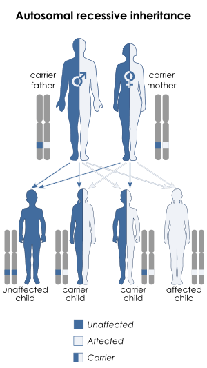
MWS is inherited in an autosomal recessive manner.[2] This means the defective gene responsible for the disorder is located on an autosome, and two copies of the defective gene (one inherited from each parent) are required in order to be born with the disorder. The parents of an individual with an autosomal recessive disorder both carry one copy of the defective gene, but usually do not experience any signs or symptoms of the disorder.
Management
The only treatment for MWS is only symptomatic, with multidisciplinary management[8]
References
- ↑ Online Mendelian Inheritance in Man (OMIM) 248700
- 1 2 Ben‐Neriah, Z.; Yagel, S.; Ariel, I. (Jul 1995). "Renal anomalies in Marden-Walker syndrome: A clue for prenatal diagnosis". American Journal of Medical Genetics. 57 (3): 417–419. doi:10.1002/ajmg.1320570310. PMID 7677143.
- 1 2 Badano JL, Mitsuma N, Beales PL, Katsanis N (2006). "The ciliopathies: an emerging class of human genetic disorders". Annu Rev Genomics Hum Genet. 7: 125–148. doi:10.1146/annurev.genom.7.080505.115610. PMID 16722803.
- ↑ https://www.orpha.net/data/patho/GB/uk-MardenWalker.pdf
- ↑ http://www.belsurg.org/uploaded_pdfs/104/104_101_103.pdf
- ↑ http://rarediseases.info.nih.gov/GARD/Condition/6973/MardenWalker_syndrome.aspx
- ↑ http://www.rarediseases.org/rare-disease-information/rare-diseases/byID/895/viewAbstract
- ↑ http://www.rightdiagnosis.com/m/marden_walker_syndrome/intro.htm
External links
- Marden-Walker syndrome at NIH's Office of Rare Diseases
- Marden Walker like syndrome at NIH's Office of Rare Diseases OMIM: 600920