Pleomorphic adenoma
| Pleomorphic adenoma | |
|---|---|
_parotid_gland.jpg) | |
| Pleomorphic adenoma consists of mixed epithelial (left) and mesenchymal cell components (right). The latter often exhibits myxofibrous appearance and in some instances shows chondromatous differentiation. | |
| Classification and external resources | |
| Specialty | oncology |
| ICD-10 | D11 |
| ICD-9-CM | 210.2 |
| ICD-O | 8940/0 |
| OMIM | 181030 |
| eMedicine | radio/531 |
| MeSH | D008949 |
Pleomorphic adenoma is a common benign salivary gland neoplasm characterised by neoplastic proliferation of parenchymatous glandular cells along with myoepithelial components, having a malignant potentiality. It is the most common type of salivary gland tumor and the most common tumor of the parotid gland. It derives its name from the architectural pleomorphism (variable appearance) seen by light microscopy. It is also known as "Mixed tumor, salivary gland type", which describes its pleomorphic appearance as opposed to its dual origin from epithelial and myoepithelial elements.
Clinical Presentation
The tumor is usually solitary and presents as a slow growing, painless, firm single nodular mass. Isolated nodules are generally outgrowths of the main nodule rather than a multinodular presentation. It is usually mobile unless found in the palate and can cause atrophy of the mandibular ramus when located in the parotid gland. When found in the parotid tail, it may present as an eversion of the ear lobe. Though it is classified as a benign tumor, pleomorphic adenomas have the capacity to grow to large proportions and may undergo malignant transformation, to form carcinoma ex-pleomorphic adenoma, a risk that increases with time (9.5% chance to convert into malignancy in 15 years). Although it is "benign" the tumor is aneuploid, it can recur after resection, it invades normal adjacent tissue and distant metastases have been reported after long (+10 years) time intervals.
Histology
Histologically, it is highly variable in appearance, even within individual tumors. Classically it is biphasic and is characterized by an admixture of polygonal epithelial and spindle-shaped myoepithelial elements in a variable background stroma that may be mucoid, myxoid, cartilaginous or hyaline. Epithelial elements may be arranged in duct-like structures, sheets, clumps and/or interlacing strands and consist of polygonal, spindle or stellate-shaped cells (hence pleiomorphism). Areas of squamous metaplasia and epithelial pearls may be present. The tumor is not enveloped, but it is surrounded by a fibrous pseudocapsule of varying thickness. The tumor extends through normal glandular parenchyma in the form of finger-like pseudopodia, but this is not a sign of malignant transformation.
The tumor often displays characteristic chromosomal translocations between chromosomes #3 and #8. This causes the PLAG gene to be juxtaposed to the gene for beta catenin. This activates the catenin pathway and leads to inappropriate cell division.
Diagnosis
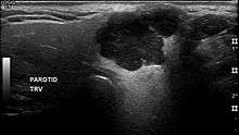
The diagnosis of salivary gland tumors utilize both tissue sampling and radiographic studies. Tissue sampling procedures include fine needle aspiration (FNA) and core needle biopsy (bigger needle comparing to FNA). Both of these procedures can be done in an outpatient setting. Diagnostic imaging techniques for salivary gland tumors include ultrasound, computer tomography (CT) and magnetic resonance imaging (MRI).
Fine needle aspiration biopsy (FNA), operated in experienced hands, can determine whether the tumor is malignant in nature with sensitivity around 90%.[1][2] FNA can also distinguish primary salivary tumor from metastatic disease.
Core needle biopsy can also be done in outpatient setting. It is more invasive but is more accurate compared to FNA with diagnostic accuracy greater than 97%.[3] Furthermore, core needle biopsy allows more accurate histological typing of the tumor.
In terms of imaging studies, ultrasound can determine and characterize superficial parotid tumors. Certain types of salivary gland tumors have certain sonographic characteristics on ultrasound.[4] Ultrasound is also frequently used to guide FNA or core needle biopsy.
CT allows direct, bilateral visualization of the salivary gland tumor and provides information about overall dimension and tissue invasion. CT is excellent for demonstrating bony invasion. MRI provides superior soft tissue delineation such as perineural invasion when compared to CT only.[5]
Treatment
Overall, the mainstay of the treatment for salivary gland tumor is surgical resection. Needle biopsy is highly recommended prior to surgery to confirm the diagnosis. More detailed surgical technique and the support for additional adjuvant radiotherapy depends on whether the tumor is malignant or benign.
Surgical treatment of parotid gland tumors is sometimes difficult, partly because of the anatomical relationship of the facial nerve and the parotid lodge, but also through the increased potential for postoperative relapse. Thus, detection of early stages of a tumor of the parotid gland is extremely important in terms of prognosis after surgery.[6]
Generally, benign tumors of the parotid gland are treated with superficial(Patey's operation) or total parotidectomy with the latter being the more commonly practiced due to high incidence of recurrence.[7] The facial nerve should be preserved whenever possible. The benign tumors of the submandibular gland is treated by simple excision with preservation of mandibular branch of the trigeminal nerve, the hypoglossal nerve, and the lingual nerve.[8] Other benign tumors of minor salivary glands are treated similarly.
Malignant salivary tumors usually require wide local resection of the primary tumor. However, if complete resection cannot be achieved, adjuvant radiotherapy should be added to improve local control.[9][10] This surgical treatment has many sequellae such as cranial nerve damage, Frey's syndrome, cosmetic problems, etc.
Usually about 44% of the patients have a complete histologic removal of the tumor and this refers to the most significant survival rate.
See also
- Warthin's tumor - monomorphic adenoma
- Carcinoma
- Sialadenitis
References
- ↑ Cohen EG, Patel SG, Lin O, et al. (Jun 2004). "Fine-needle aspiration biopsy of salivary gland lesions in a selected patient population". Arch Otolaryngol Head Neck Surg. 130 (6): 773–8. doi:10.1001/archotol.130.6.773. PMID 15210562.
- ↑ Batsakis JG, Sneige N, el-Naggar AK (Feb 1992). "Fine-needle aspiration of salivary glands: its utility and tissue effects". Ann Otol Rhinol Laryngol. 101 (2 Pt 1): 185–8. PMID 1739267.
- ↑ Wan YL, Chan SC, Chen YL, et al. (Oct 2004). "Ultrasonography-guided core-needle biopsy of parotid gland masses". AJNR Am J Neuroradiol. 25 (9): 1608–12. PMID 15502149.
- ↑ Białek EJ, Jakubowski W, Karpińska G (Sep 2003). "Role of ultrasonography in diagnosis and differentiation of pleomorphic adenomas: work in progress". Arch Otolaryngol Head Neck Surg. 129 (9): 929–33. doi:10.1001/archotol.129.9.929. PMID 12975263.
- ↑ Koyuncu M, Seşen T, Akan H, et al. (Dec 2003). "Comparison of computed tomography and magnetic resonance imaging in the diagnosis of parotid tumors". Otolaryngol Head Neck Surg. 129 (6): 726–32. doi:10.1016/j.otohns.2003.07.009. PMID 14663442.
- ↑ Alexandru Bucur; Octavian Dincă; Tiberiu Niță; Cosmin Totan; Cristian Vlădan (Mar 2011). "Parotid tumors: our experience". Rev. chir. oro-maxilo-fac. implantol. (in Romanian). 2 (1): 7–9. ISSN 2069-3850. 18. Retrieved 2012-06-06.(webpage has a translation button)
- ↑ Stennert E, Guntinas-Lichius O, Klussmann JP, Arnold G (Dec 2001). "Histopathology of pleomorphic adenoma in the parotid gland: a prospective unselected series of 100 cases". Laryngoscope. 111 (12): 2195–200. doi:10.1097/00005537-200112000-00024. PMID 11802025.
- ↑ Leonetti JP, Marzo SJ, Petruzzelli GJ, Herr B (Sep 2005). "Recurrent pleomorphic adenoma of the parotid gland". Otolaryngol Head Neck Surg. 133 (3): 319–22. doi:10.1016/j.otohns.2005.04.008. PMID 16143173.
- ↑ Ganly I, Patel SG, Coleman M, Ghossein R, Carlson D, Shah JP (Jul 2006). "Malignant minor salivary gland tumors of the larynx". Arch Otolaryngol Head Neck Surg. 132 (7): 767–70. doi:10.1001/archotol.132.7.767. PMID 16847187.
- ↑ Terhaard CH, Lubsen H, Rasch CR, et al. (Jan 2005). "The role of radiotherapy in the treatment of malignant salivary gland tumors". Int J Radiat Oncol Biol Phys. 61 (1): 103–11. doi:10.1016/j.ijrobp.2004.03.018. PMID 15629600.