Posterior superior alveolar nerve
| Posterior superior alveolar nerve | |
|---|---|
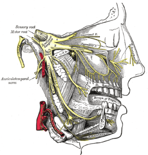 Distribution of the maxillary and mandibular nerves, and the submaxillary ganglion. (Posterior sup. alveolar labeled at center.) | |
 A sketch of the Posterior Super Nasal Nerve | |
| Details | |
| Innervates | maxillary sinus, molars, dental alveolus |
| Identifiers | |
| Latin | rami alveolares superiores posteriores nervi maxillaris |
| TA | A14.2.01.050 |
| FMA | 75545 |
The posterior superior alveolar branches (posterior superior dental branches) arise from the trunk of the maxillary nerve just before it enters the infraorbital groove; they are generally two in number, but sometimes arise by a single trunk.
They descend on the tuberosity of the maxilla and give off several twigs to the gums and neighboring parts of the mucous membrane of the cheek.
They then enter the alveolar canals on the infratemporal surface of the maxilla, and, passing from behind forward in the substance of the bone, communicate with the middle superior alveolar nerve, and give off branches to the lining membrane of the maxillary sinus and gingival and dental branches to each molar tooth from a superior dental plexus; these branches enter the apical foramina at the roots of the teeth.
The posterior superior alveolar nerve innervates the second and third maxillary molars, and two of the three roots of the maxillary first molar (all but the mesiobuccal root). When giving a posterior superior alveolar nerve block, it will anesthetize the mesialbuccal root of the maxillary first molar approximately 72% of the time.
See also
Additional images
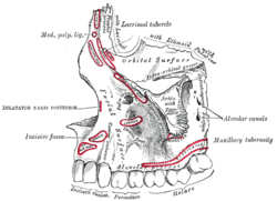 Left maxilla. Outer surface.
References
This article incorporates text in the public domain from the 20th edition of Gray's Anatomy (1918)
External links
- cranialnerves at The Anatomy Lesson by Wesley Norman (Georgetown University) (V)
- lesson9 at The Anatomy Lesson by Wesley Norman (Georgetown University) (latnasalwall4)
- MedEd at Loyola GrossAnatomy/h_n/cn/cn1/cnb2.htm