Proteus (bacterium)
| Proteus | |
|---|---|
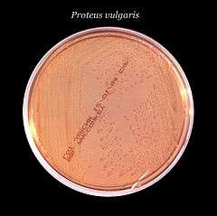 | |
| Proteus vulgaris growth in MacConkey agar culture plate | |
| Scientific classification | |
| Domain: | Bacteria |
| Phylum: | Proteobacteria |
| Class: | Gammaproteobacteria |
| Order: | Enterobacteriales |
| Family: | Enterobacteriaceae |
| Genus: | Proteus Hauser 1885 |
| Species | |
|
P. hauseri | |
Proteus is a genus of Gram-negative Proteobacteria. Proteus bacilli are widely distributed in nature as saprophytes, being found in decomposing animal matter, sewage, manure soil, and human and animal feces. They are opportunistic pathogens, commonly responsible for urinary and septic infections, often nosocomial.
Clinical significance
Three species—P. vulgaris, P. mirabilis, and P. penneri—are opportunistic human pathogens. Proteus includes pathogens responsible for many human urinary tract infections.[1] P. mirabilis causes wound and urinary tract infections. Most strains of P. mirabilis are sensitive to ampicillin and cephalosporins. P. vulgaris is not sensitive to these antibiotics. However, this organism is isolated less often in the laboratory and usually only targets immunosuppressed individuals. P. vulgaris occurs naturally in the intestines of humans and a wide variety of animals, and in manure, soil, and polluted waters. P. mirabilis, once attached to the urinary tract, infects the kidney more commonly than E. coli. P. mirabilis is often found as a free-living organism in soil and water.
About 10–15% of kidney stones are struvite stones, caused by alkalinization of the urine by the action of the urease enzyme (which splits urea into ammonia and carbon dioxide) of Proteus (and other) bacterial species.
Identification
Proteus species do not usually ferment lactose, but have shown to be capable lactose fermenters depending on the species in a triple sugar iron (TSI) test. Since it belongs to the family Enterobacteriaceae, general characters are applied on this genus. It is oxidase-negative but catalase- and nitrate-positive. Specific tests include positive urease (which is the fundamental test to differentiate Proteus from Salmonella) and phenylalanine deaminase tests.
On the species level, indole is considered reliable, as it is positive for P. vulgaris, but negative for P. mirabilis. Most strains produce a powerful urease enzyme, which rapidly hydrolyzes urea to ammonia and carbon monoxide; exceptions are some Providencia strains. Species can be motile,[2] and have characteristic "swarming" patterns.[3][4] Underlying these behaviors are the somatic O and flagellar H antigens, so named based on Kauffman–White classification. This system is based on historic observations of Edmund Weil (1879–1922) and Arthur Felix (1887–1956) of a thin surface film produced by agar-grown flagellated Proteus strains, a film that resembled the mist produced by breath on a glass. Flagellated (swarming, motile) variants were therefore designated H forms (German Hauch, for film, literally breath or mist); nonflagellated (nonswarming, nonmotile) variants growing as isolated colonies and lacking the surface film were designated as O forms (German ohne Hauch, without film [i.e., without surface film of mist droplets]).[5][6][7][8]
The cell wall O-antigen of certain strains of Proteus, such as OX-2, OX-19, OX-k, crossreact with several species of Rickettsiae. These Proteus antigens can be used in laboratory to detect the presence of antibodies against certain Rickettsiae members in patient's serum. This test is called Weil-Felix reaction after its originators.
References
| Wikimedia Commons has media related to Proteus (bacterium). |
- ↑ Guentzel MN (1996). Baron S; et al., eds. Escherichia, Klebsiella, Enterobacter, Serratia, Citrobacter, and Proteus. In: Barron's Medical Microbiology (4th ed.). Univ of Texas Medical Branch. ISBN 0-9631172-1-1. (via NCBI Bookshelf).
- ↑ Ryan KJ; Ray CG, eds. (2004). Sherris Medical Microbiology (4th ed.). McGraw Hill. ISBN 0-8385-8529-9.
- ↑ Rauprich O, Matsushita M, Weijer CJ, Siegert F, Esipov SE, Shapiro JA (November 1996). "Periodic phenomena in Proteus mirabilis swarm colony development". J. Bacteriol. 178 (22): 6525–38. PMC 178539
 . PMID 8932309.
 - ↑ Matsuyama T, Takagi Y, Nakagawa Y, Itoh H, Wakita J, Matsushita M (January 2000). "Dynamic aspects of the structured cell population in a swarming colony of Proteus mirabilis". J. Bacteriol. 182 (2): 385–93. doi:10.1128/JB.182.2.385-393.2000. PMC 94287. PMID 10629184.
- ↑ See also de:Kauffmann-White-Schema in the German Wikipedia.
- ↑ Weil, E. & Felix, A. (1917) Wien. Klin. Wschr. 30, 1509, cited in Smith, R.W. & Koffler, H., Bacterial Flagella, In Advances in Microbial Physiology, Vol. 6 (A.H. Rose & J.F. Wilkinson, Eds.), p. 251, Academic Press, 1971
- ↑ Rietschel, E.T. & Westphal, O. Endotoxin: Historical Perspectives, In Endotoxin in Health Disease (H. Brade, Ed.), p. 11, CRC Press, 1999.
- ↑ Hahon, N., Ed. Selected Papers on the Pathogenic Rickettsiae, p. 79, Harvard University Press, 1968.