Rete pegs
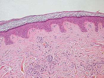
Skin epithelium (purple) with lamina propria (underlying connective tissue) (pink) -- the epithelium exhibits rete pegs.
Rete pegs (or rete processes, or rete ridges) are the epithelial extensions that project into the underlying connective tissue in both skin and mucous membranes.
In the epithelium of the mouth, the attached gingiva exhibit rete pegs, while the sulcular[1] and junctional epithelia do not.[2]
Also known as papillae, they are downward thickenings of the epidermis between the dermal papillae.
References
- ↑ Itoiz, ME; Carranza, FA: The Gingiva. In Newman, MG; Takei, HH; Carranza, FA; editors: Carranza’s Clinical Periodontology, 9th Edition. Philadelphia: W.B. Saunders Company, 2002. pages 23.
- ↑ Page, RC; Schroeder, HE. "Pathogenesis of Inflammatory Periodontal Disease: A Summary of Current Work." Lab Invest 1976;34(3):235-249
This article is issued from Wikipedia - version of the 6/29/2016. The text is available under the Creative Commons Attribution/Share Alike but additional terms may apply for the media files.