Ventricular remodeling
Ventricular remodeling (or cardiac remodeling)[1] refers to the changes in size, shape, structure, and function of the heart. This can happen as a result of exercise (physiological remodelling) or after injury to the heart muscle (pathological remodelling).[2] The injury is typically due to acute myocardial infarction (usually transmural or ST segment elevation infarction), but may be from a number of causes that result in increased pressure or volume, causing pressure overload or volume overload (forms of strain) on the heart. Chronic hypertension, congenital heart disease with intracardiac shunting, and valvular heart disease may also lead to remodeling. After the insult occurs, a series of histopathological and structural changes occur in the left ventricular myocardium that lead to progressive decline in left ventricular performance. Ultimately, ventricular remodeling may result in diminished contractile (systolic) function and reduced stroke volume.
Physiological remodelling is reversible while pathological remodelling is mostly irreversible. Remodeling of the ventricles under left/right pressure demand make mismatches inevitable. Pathologic pressure mismatches between the pulmonary and systemic circulation guide compensatory remodeling of the left and right ventricles. The term "reverse remodeling" in cardiology implies an improvement in ventricular mechanics and function following a remote injury or pathological process.
Ventricular remodeling may include ventricular hypertrophy, ventricular dilation, cardiomegaly, and other changes. It is an aspect of cardiomyopathy, of which there are many types. Concentric hypertrophy is due to pressure overload, while eccentric hypertrophy is due to volume overload.[3]
Pathophysiology
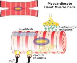
The cardiac myocyte is the major cell involved in remodeling. Fibroblasts, collagen, the interstitium, and the coronary vessels to a lesser extent, also play a role. A common scenario for remodeling is after myocardial infarction. There is myocardial necrosis (cell death) and disproportionate thinning of the heart. This thin, weakened area is unable to withstand the pressure and volume load on the heart in the same manner as the other healthy tissue. As a result, there is dilatation of the chamber arising from the infarct region. The initial remodeling phase after a myocardial infarction results in repair of the necrotic area and myocardial scarring that may, to some extent, be considered beneficial since there is an improvement in or maintenance of LV function and cardiac output. Over time, however, as the heart undergoes ongoing remodeling, it becomes less elliptical and more spherical. Ventricular mass and volume increase, which together adversely affect cardiac function. Eventually, diastolic function, or the heart's ability to relax between contractions may become impaired, further causing decline.
Evaluation
Remodeling of the heart is evaluated by performing an echocardiogram. The size and function of the atria and ventricles can be characterized using this test.
Treatment
Many factors influence the time course and extent of remodeling, including the severity of the injury, secondary events (recurrent ischemia or infarction), neurohormonal activation, genetic factors and gene expression, and treatment. Medications may attenuate remodeling. Angiotensin-converting enzyme (ACE) inhibitors have been consistently shown to decrease remodeling in animal models or transmural infarction and chronic pressure overload. Clinical trials have shown that ACE inhibitor therapy after myocardial infarction leads to improved myocardial performance, improved ejection fraction, and decreased mortality compared to patients treated with placebo. Likewise, inhibition of aldosterone, either directly or indirectly, leads to improvement in remodeling.[4] Carvedilol,a 3rd generation beta blocker, may actually reverse the remodeling process by reducing left ventricular volumes and improving systolic function.[5][6] Early correction of congenital heart defects, if appropriate, may prevent remodeling, as will treatment of chronic hypertension or valvular heart disease. Often, reverse remodeling, or improvement in left ventricular function, will also be seen.
See also
References
- ↑ Mihl, C.; Dassen, W. R. M.; Kuipers, H. (2008). "Cardiac remodelling: Concentric versus eccentric hypertrophy in strength and endurance athletes". Netherlands Heart Journal. 16 (4): 129–33. doi:10.1007/BF03086131. PMC 2300466
 . PMID 18427637.
 - ↑ Ventricular remodeling at the US National Library of Medicine Medical Subject Headings (MeSH)
- ↑ Katz, Daniel H.; Beussink, Lauren; Sauer, Andrew J.; Freed, Benjamin H.; Burke, Michael A.; Shah, Sanjiv J. (2013). "Prevalence, Clinical Characteristics, and Outcomes Associated with Eccentric Versus Concentric Left Ventricular Hypertrophy in Heart Failure with Preserved Ejection Fraction". The American Journal of Cardiology. 112 (8): 1158–64. doi:10.1016/j.amjcard.2013.05.061. PMID 23810323.
- ↑ Pitt, Bertram; Remme, Willem; Zannad, Faiez; Neaton, James; Martinez, Felipe; Roniker, Barbara; Bittman, Richard; Hurley, Steve; Kleiman, Jay; Gatlin, Marjorie (2003). "Eplerenone, a Selective Aldosterone Blocker, in Patients with Left Ventricular Dysfunction after Myocardial Infarction". New England Journal of Medicine. 348 (14): 1309–21. doi:10.1056/NEJMoa030207. PMID 12668699.
- ↑ Khattar, R. S. (2003). "Effects of ACE-inhibitors and beta-blockers on left ventricular remodeling in chronic heart failure". Minerva cardioangiologica. 51 (2): 143–54. PMID 12783070.
- ↑ "Reverse Cardiac Remodeling: A Marker of Better Prognosis in Heart Failure". Arquivos Brasileiros de Cardiologia.
Further reading
- "Left Ventricular Remodeling in Heart Failure: Current Concepts in Clinical Significance and Assessment". imaging.onlinejacc.org. Retrieved 2016-02-12.