VisualSonics
|
| |
| Industry | Biotechnology |
|---|---|
| Founded | 1999 |
| Headquarters | Toronto, Canada |
Key people | Kevin M. Goodwin, President & CEO |
| Products | Preclinical Imaging Modalities |
Number of employees | ~100 (2010) |
| Website | www.VisualSonics.com |
VisualSonics is a manufacturer of real-time, in vivo, high-resolution micro-imaging systems designed specifically for preclinical research and is a wholly owned subsidiary of SonoSite, Inc.
VisualSonics’ imaging technologies allow researchers at pharmaceutical and biotechnology companies, hospitals and universities to conduct research in cardiovascular, cancer, neurobiology and developmental biology areas. The micro-imaging technologies support research applications that include genetic research, phenotypic studies and drug development. VisualSonics high-frequency micro-imaging platforms combine high-resolution, real-time in vivo imaging with quantifiable data that have been published in over 850 scientific publications globally.
VisualSonics is based out of Toronto, Ontario, Canada with operations in more than 30 countries. European operations are conducted out of Science Park, Amsterdam, Netherlands[1] and Asia Pacific operations out of Singapore.
History

VisualSonics was founded in 1999 by medical physicist Dr. Stuart Foster,[2] who has been involved in the development of high-frequency ultrasonic systems since 1983. The company was started with support from the Canadian Institutes of Health Research (CIHR), Ontario R&D Challenge Fund (ORDCF), National Cancer Institute of Canada (NCIC), and venture capital investment.[3]
VisualSonics was acquired by SonoSite, Inc., a leading provider of hand-carried and mountable ultrasound, and impedance cardiography equipment on June 30, 2010.
Technology
VisualSonics manufactures preclinical imaging instrumentation built on its proprietary high-frequency ultrasound imaging technology.[4] High-frequency ultrasound enables high-resolution in vivo imaging of small-animal models typically used in scientific research and the preclinical stage of the drug development process.
The drug development process is broadly divided into three stages: drug discovery, preclinical studies and clinical studies. Before a particular drug can be tested on humans, its safety and efficacy must be assessed in the preclinical drug development stage.
Conventional ultrasound systems used in human (or clinical) applications operate in the 3–15 MHz frequency range, providing spatial resolution down to 300 micrometers (µm), and penetrate to a depth of 8 centimeters. These specifications are sufficient to image human organs. Conversely, when imaging a small animal such as a mouse, much higher resolution is necessary to provide useful images while depth of penetration is not required. Trading off depth penetration for higher resolution, VisualSonics systems enable imaging down to 30 µm for depths of 3 centimeters.
In addition to high-frequency ultrasound VisualSonics launched its Photoacoustic Imaging[5] platform which combines high-resolution ultrasound with the specificity of optical imaging. Photoacoustics is a non-ionizing, functional imaging modality capable of generating high contrast images of optical absorption at depths significantly greater than traditional optical imaging techniques.[6]
Products

VS40
The VS40 was the first high-frequency ultrasound commercial imaging system for animal research.[7] The system was more of a prototype device than a first-generation product, and it was sold primarily to research institutions. Even so, the VS40 was hailed by the scientific community as producing far superior images than clinical systems.[8] The VS40 system involved a single element mechanical transducer that could emit ultrasound waves in the range of 20 to 55 MHz.[9] Imaging resolution was between 60 µm and 100 µm, compared to about 300 µm for clinical systems. Only two-dimensional image acquisition was possible, of up to 4 frames per second. Thus, the Doppler function was only suitable for imaging low velocity blood flow in veins, small arteries, arterioles, or in the embryonic vasculature.

Vevo 660
Vevo 660 system was the second-generation high-frequency imaging solution launched by VisualSonics in 2003. Transducers on the system were optimized for 30 to 55 MHz operation. The Vevo 660 system offered high-resolution down to 30 μm, and the system was able to construct 3D images based on a compilation of 2D images. Image acquisition was improved to 30 frames per second, enabling imaging of fast events such as heartbeats and blood flow in small animals. Software quantification packages such as Power Doppler provided researchers with flexibility in studying blood flow and other functional information.
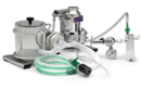
An integrated anesthesia system, and an image-guided injection system was developed to allow researchers to reduce the need for invasive surgeries when specific substances such as drugs and stem cells needed to be injected at an exact location in the animal body to allow for the most accurate results.

Vevo 770
The Vevo 770 micro-ultrasound system was released in 2006.[10] Specific improvements included, frame rate enhancements to 100 frames per second, compared with 30 in the Vevo 660 system.[11] The higher frame rates allowed researchers to visualize fast changing valvular and myocardial heart functions in small animals (mice heartbeat etc.). Furthermore, improved electronics and signal detection allowed image penetration depth to increase by about 25%. To support the need for data analysis and quantification of images acquired by the ultrasound machines, numerous software modes were offered on the Vevo 770 system. New features on the Vevo 770 system include Power Doppler Mode, a mode that allows for the visualization and quantification of relative microvascularity in vivo for anti-angiogenic studies. In addition, Advanced Cardiovascular Functionality provides Tissue Doppler Imaging (TDI), Integrated Blood Pressure Measurement, Anatomical M-Mode, Automated Left Ventricular Quantification, and Advanced Measurements and Annotation Functionality.
In addition, an integrated animal platform was also released, allowing the researcher to monitor in real-time animal physiology such as heart rate, temperature rate, respiration rate, and blood pressure while they are being imaged by ultrasound.


Vevo 2100
The fourth-generation Vevo 2100 system released in 2008 offered high-frequency 256-element linear array technology versus the single-element transducers of previous products.[12] This is not only the world's first array transducer technology to be released for preclinical imaging,[13] but also significantly increased transducer reliability, and the quality of images produced. Frame rates were improved to 300-400 frames per second, and up to 1000 frames per second in a narrow field of view.
Many software enhancements were also offered, such as Color Doppler Mode for visualizing direction of blood flow, and VevoStrain software to quantify advanced myocardial functions. Vevo 2100 platform continues to be advanced with newer imaging modes such as 3D Color Doppler Mode, Cancer imaging functionality - Nonlinear Contrast Mode, Perfusion quantification with VevoCQ Software, ECG-Gated Acquisition and digital-RF functionality.
The system supports exporting of datasets in various open formats for study management and longitudinal analysis.
Vevo LAZR Photoacoustics Imaging System
The Vevo LAZR Photoacoustics Imaging System was released April 2011 at the AACR 2011.
Vevo 3100
The Vevo 3100 system was released in September 2014 at the World Molecular Imaging Congress (WMIC).[14] The fifth-generation micro-ultrasound product introduced a touch based user interface and a form factor with a reduced footprint and weight compared to the previous Vevo 2100. Software enhancements include the introduction of Vevo HD Imaging Technology, a software based real-time image processing algorithm that reduces speckle noise and artifacts.[15]
A new line of ultra-high frequency linear array transducers referred to as the MX Transducers were released exclusively for the Vevo 3100 imaging platform. The MX Transducers are lightweight and ergonomic improvements over the earlier MS (MicroScan) Transducers.[14]
Vevo MD - Clinical
Vevo MD was approved by the FDA for clinical use in April 2016.[16] European Regulatory approval was granted in January 2016 and marked Visualsonics' introduction into the clinical market. Vevo MD is the first Ultra High Frequency (UHF) clinical ultrasound system.[17][18] The UHF line of transducers with frequencies up to 70 MHz were released specifically for the system's use in the clinical space. The Vevo MD is built on the Vevo 3100 imaging platform in which the two products share similar form factors and styling. The touch based user interface introduced for the Vevo 3100 is also present on the Vevo MD clinical system.
Applications
Preclinical small animal studies are a key part of medical and biological research. The non-invasive high-resolution ultrasound technology represented in the VisualSonics product line represents a significant improvement in the quality and speed with which information about disease models may be obtained. Furthermore, the use of non-invasive imaging has the potential to significantly reduce the number of animals required for research, by eliminating the need for histological approaches in some longitudinal animal studies.
Ultrasound has been a common imaging modality in cardiovascular research and the availability of high-resolution has meant that murine models of cardiovascular disease may be imaged with some of the same techniques used in larger animals and humans. This access to high quality cardiovascular data in mice has facilitated better research into human cardiovascular disease, medical and surgical treatments, and improved the lives of millions of people. Ultrasound is less common in cancer research. However, the advent of high-resolution ultrasound provides the opportunity to image both subcutaneous and orthotopic tumors easily, quickly and with higher accuracy and precision than other modalities in small animal disease models.
The application of high-resolution ultrasound in preclinical development opens the possibility of using ultrasound more frequently as a component of clinical trials by translating the techniques developed in the animal models directly into the clinic. The key areas of high-frequency ultrasound include:
- Molecular Imaging
- Cardiovascular Research
- Neurobiology
- Developmental Biology
- Cancer Research
- Nephrology
- Toxicology
- Gene Delivery
- Image-Guided Injections
Awards and recognitions
In 2005, VisualSonics’ President and CEO, Mr. Tom Little, received Ernst & Young’s “Entrepreneur of the YearTM 2005 Ontario Region Health Science Award” based on the large growth he brought to VisualSonics in a few years.[19]
In 2006, VisualSonic’s Founder and CSO, Dr. Stuart Foster, received the 2006 Ernest C. Manning Award Foundation’s “Award of Distinction” in recognition of his innovative concept of micro-ultrasound and its contribution to the field of pre-clinical imaging.[20]
In 2007, VisualSonics was awarded the “Best Customer Value” Award by Frost & Sullivan for its Vevo 770 system.[19] Frost & Sullivan is a global growth consulting company conducting extensive primary and secondary research including hundreds of customer and subject matter expert interviews. This was compiled in the “North American Preclinical Small Animal Imaging Markets” report, which recognized the Vevo systems by VisualSonics as having an extremely satisfied and rapidly growing customer base in both academic research and industry.
References
- ↑ "VisualSonics - NFIA". NFIA. Retrieved 2016-06-04.
- ↑ "Dr Stuart Foster, PhD (Scientist Profile), Sunnybrook Research Institute, Toronto, Canada". Retrieved 2012-05-01.
- ↑ "Sunnybrook: A Sound Venture". Retrieved 2010-02-05.
- ↑ "Toronto firm pioneers high-frequency ultrasound, The Star (July 2010)". 2010-07-27. Retrieved 2010-07-27.
- ↑ "Photoacoustic Imaging. The sound of light, The Economist (Sept 2009)". 2009-06-04. Retrieved 2009-09-01.
- ↑ "Biomedical photoacoustics beyond thermal expansion using triggered nanodroplet vaporization for contrast-enhanced imaging, Pubmed.gov(Jan 2012)". Retrieved 2012-01-01.
- ↑ "FBO Daily: VS40". Retrieved 2010-02-08.
- ↑ Zhou YQ, Foster FS, Qu DW, Zhang M, Harasiewicz KA, Adamson SL (August 2002). "Applications for multifrequency ultrasound biomicroscopy in mice from implantation to adulthood". Physiological Genomics. 10 (2): 113–26. doi:10.1152/physiolgenomics.00119.2001. PMID 12181368.
- ↑ Foster FS, Zhang MY, Zhou YQ, et al. (September 2002). "A new ultrasound instrument for in vivo microimaging of mice". Ultrasound in Medicine & Biology. 28 (9): 1165–72. doi:10.1016/S0301-5629(02)00567-7. PMID 12401387.
- ↑ "MedGadget: Vevo 770 Micro-imaging System". Retrieved 2010-02-08.
- ↑ "VisualSonics: Vevo 770 Micro-imaging System". Retrieved 2010-02-08.
- ↑ "VisualSonics: Vevo 2100 Micro-imaging System". Retrieved 2010-02-08.
- ↑ "HighBeam: Visualsonics introduces World's First high Frequency Ultrasound Digital Platform with Array Transducer Technology; The New Vevo 2100 Micro Imaging Platform". Retrieved 2010-02-08.
- 1 2 "FUJIFILM VisualSonics Unveils the Ultimate Pre-Clinical Imaging Experience with the Vevo® 3100 at the World Molecular Imaging Congress Annual Meeting". MarketWatch. Retrieved 2016-06-04.
- ↑ "Vevo® 3100 - High-frequency ultrasound imaging for preclinical research | VisualSonics". www.visualsonics.com. Retrieved 2016-06-04.
- ↑ "Vevo MD, World's First Clinical Ultra High Frequency Ultrasound FDA Cleared |". Medgadget. 2016-04-22. Retrieved 2016-06-04.
- ↑ "FUJIFILM VisualSonics Announces CE Mark for Vevo® MD | Business Wire". www.businesswire.com. Retrieved 2016-06-04.
- ↑ "FUJIFILM Visualsonics - Vevo MD". FUJIFILM Visualsonics. Retrieved 2016-06-04.
- 1 2 "Canadian Business: One-on-One with Tom Little, President & CEO, VisualSonics". Retrieved 2016-06-04.
- ↑ "Manning Innovation Awards". Retrieved 2016-06-04.