Cat anatomy

The anatomy of the domestic cat is similar to that of other members of the genus Felis.
Mouth
 A yawning cat, exposing its mouth |
Cats have highly specialized teeth for the killing of prey and the tearing of meat. The premolar and first molar, together the carnassial pair are located on each side of the mouth. These teeth efficiently function to shear meat like a pair of scissors. While this is present in canids, it is highly developed in felines.
The cat's tongue has sharp spines, or papillae, useful for retaining and ripping flesh from a carcass. These papillae are small backward-facing hooks that contain keratin which also assist in their groom.
The cat's oral structures provide for a variety of vocalizations used for communication, including meowing, purring, hissing, growling, squeaking, chirping, clicking, and grunting.
Ears
 A cat's ear which has special fur for sensing and protection |
Thirty-two individual muscles in each ear allow for a manner of directional hearing;[1] a cat can move each ear independently of the other. Because of this mobility, a cat can move its body in one direction and point its ears in another direction. Most cats have straight ears pointing upward. Unlike dogs, flap-eared breeds are extremely rare (Scottish Folds are one such exceptional mutation). When angry or frightened, a cat will lay back its ears to accompany the growling or hissing sounds it makes. Cats also turn their ears back when they are playing or to listen to a sound coming from behind them. The fold of skin forming a pouch on the lower posterior part of the ear, known as Henry's pocket, is usually prominent in a cat's ear.[2] It is of unknown function, though it may assist in filtering sounds.
Nose

Cats are highly territorial and secreting odors plays a major role in cat communication. The nose helps cats to identify territories, other cats and mates, to locate food, and for various other causes.[3] A cat's sense of smell is believed to be about fourteen times stronger than that of humans. The rhinarium (the leathery bit of nose we see) is quite tough to allow it to absorb rather rough treatment sometimes. The color varies according to the genotype (genetic makeup) of the cat. Cat's skin has the same color as the fur but the color of the nose leather is probably dictated by a dedicated gene. Cats with white fur have skin susceptible to damage by ultraviolet light that may cause cancer. Extra care is required when outside in hot sun.[4]
Legs
Cats, like dogs, are digitigrades. They walk directly on their toes, with the bones of their feet making up the lower part of the visible leg.[5] Domestic cats are capable of walking very precisely, as is true for all cats. Like all felines, they directly register; that is, they place each hind paw (almost) directly in the print of the corresponding forepaw, minimizing noise and visible tracks. This also provides sure footing for their hind paws when they navigate rough terrain. The two back legs allow falling and leaping far distances without injury.
Unlike most mammals, when cats walk, they use a "pacing" gait; that is, they move the two legs on one side of the body before the legs on the other side. This trait is shared with camels and giraffes. As a walk speeds up into a trot, a cat's gait will change to be a "diagonal" gait, similar to that of most other mammals: the diagonally opposite hind and forelegs will move simultaneously. Cat height can vary depending on breed and/or gender, but is usually around 12 inches or 30.5 centimeters[6]
Claws
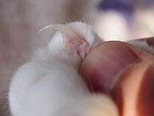
Like nearly all members of the family Felidae, cats have retractable claws. In their normal, relaxed position, the claws are sheathed with the skin and fur around the toe pads. This keeps the claws sharp by preventing wear from contact with the ground and allows the silent stalking of prey. The claws on the forefeet are typically sharper than those on the hind feet.[7] Cats can voluntarily extend their claws on one or more paws. They may extend their claws in hunting or self-defense, climbing, "kneading", or for extra traction on soft surfaces (bedspreads, thick rugs, skin, etc.). It is also possible to make a cooperative cat extend its claws by carefully pressing both the top and bottom of the paw. The curved claws can become entangled in carpet or thick fabric, which can cause injury if the cat is unable to free itself.
Most cats have five claws on their front paws, and four or five on their rear paws.[8] Because of an ancient mutation, however, some domestic and feral cats are born with polydactylyism (particularly in the east coast of Canada and northeast coast of the United States), with six or seven toes.
Temperature and heart rate
The normal body temperature of a cat is between 38.33 and 39.0 °C (101.0 and 102.2 °F).[9] A cat is considered febrile (hyperthermic) if it has a temperature of 39.5 °C (103.1 °F) or greater, or hypothermic if less than 37.5 °C (99.5 °F). For comparison, humans have an average body temperature of about 37.0 °C (98.6 °F).[10] A domestic cat's normal heart rate ranges from 140 to 220 beats per minute (bpm), and is largely dependent on how excited the cat is. For a cat at rest, the average heart rate usually is between 150 and 180 bpm, about twice that of a human, which averages 70 bpm.[11]
Skin
Cats possess rather loose skin; this allows them to turn and confront a predator or another cat in a fight, even when it has a grip on them. This is also an advantage for veterinary purposes, as it simplifies injections.[12] In fact, the lives of cats with kidney failure can sometimes be extended for years by the regular injection of large volumes of fluid subcutaneously, which serves as an alternative to dialysis.[13][14]
- Scruff
The particularly loose skin at the back of the neck is known as the scruff, and is the area by which a mother cat grips her kittens to carry them. As a result, cats tend to become quiet and passive when gripped there. This behavior also extends into adulthood, when a male will grab the female by the scruff to immobilize her while he mounts, and to prevent her from running away as the mating process takes place.[15]
This technique can be useful when attempting to treat or move an uncooperative cat. However, since an adult cat is heavier than a kitten, a pet cat should never be carried by the scruff, but should instead have its weight supported at the rump and hind legs, and at the chest and front paws.
- Primordial pouches
Some cats share common traits due to heredity. One of those is the primordial pouch, sometimes referred to as "spay sway" by owners who notice it once the cat has been spayed or neutered. It is located on a cat's belly. Its appearance is similar to a loose flap of skin that might occur if the cat had been overweight and had then lost weight. It provides a little extra protection against kicks, which are common during cat fights as a cat will try to rake with its rear claws. In wild cats, the ancestors of domesticated felines, this pouch appears to be present to provide extra room in case the animal has the opportunity to eat a large meal and the stomach needs to expand. This stomach pouch also allows the cat to bend and expand, allowing for faster running and higher jumping.[16]
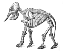
Skeleton

A – Cervical or Neck Bones (7 in number). B – Dorsal or Thoracic Bones (13 in number, each bearing a rib). C – Lumbar Bones (7 in number).D – Sacral Bones (3 in number).E – Caudal or Tail Bones (19 to 21 in number).
1 – Cranium, or Skull.
2 – Mandible, or Lower jaw.
3 – Scapula, or Shoulder-blade.
4 – Sternum, or Breast-bone.
5 – Humerus.
6 – Radius.
7 – Phalanges of the Toes.
8 – Metacarpal Bones.
9 – Carpal or Wrist-bones.
10 – Ulna.
11 – Ribs.
12 – Patella, or Knee-cap.
13 – Tibia.
14 – Metatarsal Bones.
15 – Tarsal Bones.
16 – Fibula.
17 – Femur, or Thigh-bone.
18 – Pelvis, or Hip-bone.

Cats have seven cervical vertebrae like almost all mammals, thirteen thoracic vertebrae (humans have twelve), seven lumbar vertebrae (humans have five), three sacral vertebrae (humans have five because of their bipedal posture), and, except for Manx cats and other shorter tailed cats, twenty-two or twenty-three caudal vertebrae (humans have three to five, fused into an internal coccyx). The extra lumbar and thoracic vertebrae account for the cat's enhanced spinal mobility and flexibility, compared to humans. The caudal vertebrae form the tail, used by the cat as a counterbalance to the body during quick movements. Between their vertebrae, they have elastic discs, useful for cushioning the jump landings.
Unlike human arms, cat forelimbs are attached to the shoulder by free-floating clavicle bones, which allows them to pass their body through any space into which they can fit their heads.[17]
Skull
The cat skull is unusual among mammals in having very large eye sockets and a powerful and specialized jaw.[18]:35 Compared to other felines, domestic cats have narrowly spaced canine teeth, adapted to their preferred prey of small rodents.[19]
Muscles
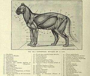
Internal Abdominal Oblique
This muscle's origin is the lumbodorsal fascia and ribs. Its insertion is at the pubis and linea alba (via aponeurosis), and its action is the compression of abdominal contents. It also laterally flexes and rotates the vertebral column.
Transversus Abdominis
This muscle is the innermost abdominal muscle. Its origin is the second sheet of the lumbodorsal fascia and the pelvic girdle and its insertion is the linea alba. Its action is the compression of the abdomen.
Rectus Abdominis
To see this muscle, first remove the extensive aponeurosis situated on the ventral surface of the cat. Its fibers are extremely longitudinal, on each side of the linea alba. It is also traversed by the inscriptiones tendinae, or what others called myosepta.
Deltoid
The deltoid muscles lie just lateral to the trapezius muscles, originating from several fibers spanning the clavicle and scapula, converging to insert at the humerus. Anatomically, there are only two deltoids in the cat, the acromiodeltoid and the spinodeltoid. However, to conform to human anatomy standards, the clavobrachialis is now also considered a deltoid and is commonly referred to as the clavodeltoid.
Acromiodeltoid
The acromiodeltoid is the shortest of the deltoid muscles. It lies lateral to (to the side of) the clavodeltoid, and in a more husky cat it can only be seen by lifting or reflecting the clavodeltoid. It originates at the acromion process and inserts at the deltoid ridge. When contracted, it raises and rotates the humerus outward.
Spinodeltoid
A stout and short muscle lying posterior to the acromiodeltoid. It lies along the lower border of the scapula, and it passes through the upper arm, across the upper end of muscles of the upper arm. It originates at the spine of the scapula and inserts at the deltoid ridge. Its action is to raise and rotate the humerus outward.
Head
Masseter
The Masseter is a great, powerful, and very thick muscle covered by a tough, shining fascia lying ventral to the zygomatic arch, which is its origin. It inserts into the posterior half of the lateral surface of the mandible. Its action is the elevation of the mandible (closing of the jaw).
Temporalis
The temporalis is a great mass of mandibular muscle, and is also covered by a tough and shiny fascia. It lies dorsal to the zygomatic arch and fills the temporal fossa of the skull. It arises from the side of the skull and inserts into the coronoid process of the mandible. It too, elevates the jaw.
Integumental
The two main integumentary muscles of a cat are the platysma and the cutaneous maximus. The cutaneous maximus covers the dorsal region of the cat and allows it to shake its skin. The platysma covers the neck and allows the cat to stretch the skin over the pectoralis major and deltoid muscles.
Neck and back
Rhomboideus
The Rhomboideus is a thick, large muscle below the Trapezius muscles. It extends from the vertebral border of the scapula to the mid-dorsal line. Its origin is from the neural spines of the first four thoracic vertebrae, and its insertion is at the vertebral border of the scapula. Its action is to draw the scapula to the dorsal.
Rhomboideus Capitis
The Rhomboideus capitis is the most cranial of the deeper muscles. It is underneath the Clavotrapezius. Its origin is the superior nuchal line, and its insertion is at the scapula. Action draws scapula cranially.
Splenius
The Splenius is the most superficial of all the deep muscles. It is a thin, broad sheet of muscle underneath the Clavotrapezius and deflecting it. It is crossed also by the Rhomboideus capitis. Its origin is the mid-dorsal line of the neck and fascia. The insertion is the superior nuchal line and atlas. It raises or turns the head.
Serratus Ventralis
The Serratus Ventralis is exposed by cutting the wing-like Latissimus Dorsi. The said muscle is covered entirely by adipose tissue. The origin is from the first nine or ten ribs and from part of the cervical vertebrae.
Serratus Dorsalis
The Serratus Dorsalis is medial to both the scapula and the Serratus Ventralis. Its origin is via apoeurosis following the length of the mid-dorsal line, and its insertion is the dorsal portion of the last ribs. Its action is to depress and retracts the ribs during breathing.
Intercostals
The Intercostals are a set of muscles sandwiched among the ribs. They interconnect ribs, and are therefore the primary respiratory skeletal muscles. They are divided into the external and the internal subscapularis. The origin and insertion are in the ribs. The intercostals pull the ribs backwards or forwards.
Caudofemoralis
The Caudofemoralis is a muscle found in the pelvic limb and is unique to the felids (cats).[20] The Caudofemoralis acts to flex the tail laterally to its respective side when the pelvic limb is bearing weight. When the pelvic limb is lifted off the ground, contraction of the Caudofemoralis causes the limb to abduct and the shank to extend by extending the hip joint.
Pectoral
Pectoantebrachialis
Pectoantebrachialis muscle is just one-half inch wide, and is the most superficial in the pectoral muscles. Its origin is the manubrium of the sternum, and its insertion is in a flat tendon on the fascia of the proximal end of the ulna. Its action is to draw the arm towards the chest. There is no human equivalent.
Pectoralis Major
The pectoralis major, also called pectoralis superficialis, is a broad triangular portion of the pectoralis muscle which is immediately below the pectoantebrachialis. It is smaller than the pectoralis minor muscle. Its origin is the sternum and median ventral raphe, and its insertion is at the humerus. Its action is to draw the arm towards the chest.
Pectoralis Minor
The pectoralis minor muscle is larger than the Pectoralis major. However, most of its anterior border is covered by the pectoralis major. Its origins are ribs three–five, and its insertion is the coracoid process of the scapula. Its actions are the tipping of the scapula and the elevation of ribs three–five.
Xiphihumeralis
The most posterior, flat, thin, and long strip of pectoral muscle is the Xiphihumeralis. It is a band of parallel fibers that is found in felines but not in humans. Its origin is the Xiphoid Process of the sternum. The insertion is the humerus.
Trapezius
In the cat there are three thin flat muscles that cover the back, and to a lesser extent, the neck. They pull the scapula toward the mid-dorsal line, anteriorly, and posteriorly.
Clavotrapezius
The most anterior of the trapezius muscles, it is also the largest. Its fibers run obliquely to the ventral surface. Its origin is the superior nuchal line and median dorsal line and its insertion is the clavicle. Its action is to draw the clavicle dorsally and towards the head.
Acromiotrapezius
Acromiotrapezius is the middle trapezius muscle. It covers the dorsal and lateral surfaces of the scapula. Its origin is the neural spines of the cervical vertebrae and its insertion is in the metacromion process and fascia of the clavotrapezius. Its action is to draw the scapula to the dorsal, and hold the two scapula together.
Spinotrapezius
Spinotrapezius, also called thoracic trapezius, is the most posterior of the three. It is triangular shaped. Posterior to the acromiotrapezius and overlaps latissimus dorsi on the front. Its origin is the neural spines of the thoracic vertebrae and its insertion is the scapular fascia. Its action is to draw the scapula to the dorsal and caudal region.
Genitalia

Female genitalia
In the female cat, the genitalia include two gonads, the uterus, the vagina, the genital passages and teats. Together with the vulva, the vagina of cat is involved in mating and provides a channel for newborns during parturition, or birth. The vagina is long and wide.[21] Genital passages are the oviducts of the cat. They are short, narrow, and not very sinuous.[21]
Male genitalia
In the male cat, the genitalia includes the penis, which is covered with small spines.[22]
See also
References
- ↑ "At Home: Care / Health: Understanding Cats". Archived from the original on 1 February 2008. Retrieved 15 August 2005.
- ↑ August, John (2009). Consultations in Feline Internal Medicine, Volume 6. Elsevier Health Sciences.
- ↑ Syufy F. "The Nose Knows Cats' Amazing Sense of Scent". About.com.
- ↑ "Cat Anatomy". cat-chitchat.pictures-of-cats.org. 9 July 2008.
- ↑ Lacquaniti, F.; Grasso, R.; Zago, M. (1 August 1999). "Motor Patterns in Walking". News Physiol. Sci. 14 (4): 168–174. PMID 11390844.
- ↑ Christensen, Wendy (2004). Outwitting Cats. Globe Pequot. p. 23. ISBN 1-59228-240-7.
- ↑ Armes, Annetta F. (22 December 1900). "Outline of Cat Lessons". The School Journal. LXI: 659. Retrieved 12 November 2007.
- ↑ Danforth, C. H. (1947). "Heredity of Polydactyly in the Cat" (PDF). Journal of Heredity. 38 (4): 107–112. PMID 20242531.
- ↑ "Normal Values For Dog and Cat Temperature, Blood Tests, Urine and other information in ThePetCenter.com". Archived from the original on 13 March 2005. Retrieved 1 August 2005.
- ↑ http://www.health.harvard.edu/press_releases/normal_body_temperature
- ↑ Cat Health And Cat Metabolism Information For The Best Cat Care. Highlander Pet Center
- ↑ "Vaccinate Your Cat at Home". Retrieved 18 October 2006.
- ↑ Kellman, Rich. "The Cat Comes Back". Retrieved 1 March 2010.
- ↑ "How to Give Subcutaneous Fluids to a Cat". wikihow.com. Retrieved 18 October 2006.
- ↑ "Scruffing your dog or cat". pets.c. Retrieved 26 February 2008.
- ↑ What Is the Primordial Pouch in Cats? by Nicholas DeMarino at pets.thenest.com
- ↑ Gillis, Rick (ed.) (22 July 2002). "Cat Skeleton". Zoolab: A Website for Animal Biology. La Crosse, WI: University of Wisconsin. Archived from the original on 6 December 2006. Retrieved 7 September 2012.
- ↑ Case, Linda P. (2003). The Cat: Its Behavior, Nutrition, and Health. Ames, IA: Iowa State University Press. ISBN 0-8138-0331-4.
- ↑ Smith, Patricia; Tchernov, Eitan (1992). Structure, Function and Evolution of teeth. Freund Publishing House Ltd. p. 217. ISBN 965-222-270-4.
- ↑ Rosenzweig, L. J. (1990). Anatomy of the Cat: Text and Dissection Guide. Wm. C. Brown Publishers Dubuque, IA. p. 110, ISBN 0697055795.
- 1 2 The cat's genital system and reproduction. aniwa.com
- ↑ Heide Schatten; Gheorghe M. Constantinescu (21 March 2008). Comparative Reproductive Biology. John Wiley & Sons. ISBN 978-0-470-39025-2.
External links
| Wikimedia Commons has media related to Cat anatomy. |
- The cat; an introduction to the study of backboned animals, especially mammals (1881)
- A laboratory guide for the dissection of the cat: An introduction to the study of anatomy (1895)
- Anatomy of the cat (1902)
Anatomy and morphology | ||
|---|---|---|
| Fields |
|  |
| Bacteria | ||
| Protists |
| |
| Plants |
| |
| Invertebrates | ||
| Mammals | ||
| Other vertebrates | ||
| Other topics | ||