Nodular fasciitis
| Nodular fasciitis | |
|---|---|
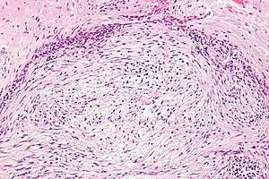 | |
| Micrograph of nodular fasciitis showing the haphazard arrangement of cells (tissue culture-like pattern). H&E stain. | |
| Classification and external resources | |
| Specialty | rheumatology |
| ICD-10 | M72.4 |
| ICD-9-CM | 728.79 |
Nodular fasciitis, also known as nodular pseudosarcomatous fasciits,[1] pseudosarcomatous fasciitis,[2] and subcutaneous pseudosarcomatous fibromatosis,[1]:992 is a benign[3] soft tissue lesion most commonly found in the superficial fascia. The lesion commonly occurs in the first three decades of life. Upper extremities and trunk are the most common affected anatomical sites. Previous history of trauma may be present. Clinically and histologically, nodular fasciitis may be mistaken for a sarcoma.
Etiology and clinical course
Until recently, nodular fasciitis have been considered a reactive process of uncertain etiology.[4] However, recent findings indicate that nodular fasciitis is a self-limited clonal neoplastic process (see below). Clinically, nodular fasciitis presents as a subcutaneous "growth" over a period of 3–6 weeks that eventually regresses. The lesion usually reaches a size of 2–3 cm. Larger lesions are unusual. Local recurrence has been described after simple surgical excision but it is rare.
Histology
- Histologically vast array of patterns.
- Short S-shaped fascicles, inflammation, accelerated mitotic index with normal mitoses.
- Essentially spindle cell proliferation.
- Stroma is rich in collagen and/or myxoid ground substance.
Additional images
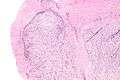 Low mag.
 Intermed. mag.
.jpg) Goldner, 400x
Goldner, 400x
See also
References
- 1 2 Freedberg, et al. (2003). Fitzpatrick's Dermatology in General Medicine. (6th ed.). McGraw-Hill. ISBN 0-07-138076-0.
- ↑ Rapini, Ronald P.; Bolognia, Jean L.; Jorizzo, Joseph L. (2007). Dermatology: 2-Volume Set. St. Louis: Mosby. ISBN 1-4160-2999-0.
- ↑ Sailon AM, Cappuccino G, Hameed M, Fleegler EJ (2008). "Nodular fasciitis of the hand over the metacarpophalangeal joint: a case report". Eplasty. 8: e38. PMC 2491338
 . PMID 18725954.
 - ↑ Oliveira AM, Chou MM. USP6-induced neoplasms: the biologic spectrum of aneurysmal bone cyst and nodular fasciitis. Hum Pathol. 2013 Jun 11. doi:pii: S0046-8177(13)00116-0. doi:10.1016/j.humpath.2013.03.005. PMID 23769422.