Surgical margin
Surgical margin, also known as tumor free margin, free margin, normal skin margin, and normal tissue margin, usually refers to the visible normal tissue or skin margin that is removed with the surgical excision of a tumor, growth, or malignancy.
Definition
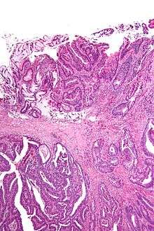
Surgical margin in a surgery report defines the visible margin or free edge of "normal" tissue seen by the surgeon with the naked eye. Surgical margin as read in a pathology report defines the histological measurement of normal or unaffected tissue surrounding the visible tumor under a microscope on a glass mounted histology section.[1][2] A "narrow" surgical margin implies that the tumor exists very close to the surgical margin, and a "wide" surgical margin implies the tumor exists far from the cut edge or the surgical margin. Narrow surgical margin using the bread loafing technique suggests that residual cancer might be left due to false negative error. A surgeon often will perform a second surgery if a narrow surgical margin is noted on a pathology report.
Associated errors and recurrence rate
This determination is made with the full understanding of "false negative error" intrinsic in the bread loafing technique of histology (also known as POMA - a term used by the NCCN).[3] The higher the false negative error is, the higher the recurrence rate of a cancer or tumor at the surgical margin. This is due to the misreading of a pathology specimen as being clear of residual tumor, when there is actually residual tumor left where the specimen was not cut and mounted on the histology slide. The "false negative error" is very low in the CCPDMA method of histology processing, and can be very high in the bread loafing (POMA) method of histology processing.[3] In the bread loafing method of processing, one will note a high false negative error rate with narrow surgical margin; and one will note a low false negative error with a wide surgical margin[4] Surgical margin has a much less significant effect on the false negative error rate of CCPDMA methods, allowing the surgeon to routinely use very narrow surgical margins (1 to 2 mm for non-melanoma skin cancer).[4]
References
- ↑ Maloney, ME., et al. Surgical Dermatopathology. Blackwell Science, 1999. pp. 107-121.
- ↑ Maxwell, JH; Thompson, LD; Brandwein-Gensler, MS; Weiss, BG; Canis, M; Purgina, B; Prabhu, AV; Lai, C; Shuai, Y; Carroll, WR; Morlandt, A; Duvvuri, U; Kim, S; Johnson, JT; Ferris, RL; Seethala, R; Chiosea, SI (1 December 2015). "Early Oral Tongue Squamous Cell Carcinoma: Sampling of Margins From Tumor Bed and Worse Local Control.". JAMA otolaryngology-- head & neck surgery. 141 (12): 1104–10. doi:10.1001/jamaoto.2015.1351. PMID 26225798.
- 1 2 Tri-Service General Hospital
- 1 2 Kimyai-Asadi, Arash; Katz, Tracy; Goldberg, Leonard H.; Ayala, Gabriel B.; Wang, Steven Q.; Vujevich, Justin J.; Jih, Ming H. (2007). "Margin Involvement after the Excision of Melanoma in Situ: The Need for Complete En Face Examination of the Surgical Margins". Dermatologic Surgery. 33 (12): 1434–9; discussion 1439–41. doi:10.1111/j.1524-4725.2007.33313.x. PMID 18076608.
Further reading
- Upile, T.; Fisher, C.; Jerjes, W.; El Maaytah, M.; Searle, A.; Archer, D.; Michaels, L.; Rhys-Evans, P.; Hopper, C.; Howard, D.; Wright, A. (2007). "The uncertainty of the surgical margin in the treatment of head and neck cancer". Oral Oncology. 43 (4): 321. doi:10.1016/j.oraloncology.2006.08.002. PMID 17112772.